How Stem Cells Decide Their Fate

Source: Memorial Sloan Kettering - On Cancer
Date: 08/13/2019
Link to original
Image of article

Stem cells are defined by their ability to differentiate into other, more specialized cell types. When one stem cell divides into two (which are then called daughter cells), three things can happen to the new cells: Both cells can continue being stem cells, both cells can differentiate into a new cell type, or the cells can go their separate ways, with one maintaining the properties of a stem cell and the other becoming something new.

“It may be surprising, but as much as stem cells have been studied, we don’t know much about how they make this commitment when they divide,” says Michael Kharas, who is in the Sloan Kettering Institute’s Molecular Pharmacology Program. Dr. Kharas led a team from SKI, in collaboration with investigators at Weill Cornell Medical College, that discovered new details about how dividing stem cells choose what to become. Their findings were published August 13 in Cell Reports.

Dr. Kharas’s lab uses human and mouse blood (hematopoietic) stem cells to study how certain types of leukemia develop.

Knocking Out an Important Protein

When both daughter cells are the same cell type, this is called symmetric division. That’s true whether they continue being stem cells or become something new. When one cell stays a stem cell and the other differentiates, it’s called asymmetric division. This happens about 30 percent of the time.

“The beauty of asymmetric division is that you’re maintaining your stem cell numbers,” Dr. Kharas explains. “If every time a stem cell divides, it loses its identity as a stem cell, you will eventually deplete all the stem cells. But if all the cells remain stem cells, you’ll never get the variety of cell types you need to form an entire blood system.”

Previous work in the Kharas lab has focused on a protein called MUSASHI-2. The researchers found that when MUSASHI-2 is knocked out in blood stem cells, the cells lose their “stemness.” They are more likely to differentiate and become nonstem cells. Additionally, they have less of the asymmetric type of division.Targeting RNA-binding proteins has been identified as a new approach in the development of drugs for leukemia.

In the new study, the investigators — led by first authors Yuanming Cheng and Hanzhi Luo, of Dr. Kharas’s lab, and Franco Izzo from Weill Cornell — focused on a different protein: METTL3. Both MUSASHI-2 and METTL3 are RNA-binding proteins, but the METTL3 protein is the key enzyme that can add chemicals called methyl groups to specific RNA nucleotides. This process is called methylation, and it decorates RNA with marks called m6A. 

Ultimately, this helps regulate the stability of the RNAs and the efficiency of protein production. Targeting RNA-binding proteins has been identified as a new approach in the development of drugs for leukemia.

Taking a Closer Look at Stem Cells

To study the role of m6A, the researchers compared the blood-forming systems of mice that had METTL3 knocked out and those that did not, to see how blood development was affected. They found that the mice without the protein seemed to have an accumulation of blood stem cells, suggesting that the cells were unable to become more specialized cells. 

To get a better look at what was going on, the lab of Dan Landau at Weill Cornell studied the cells with a type of analysis called single-cell RNA sequencing (RNA seq). RNA seq enables researchers to determine which genes are being expressed, or turned on, in cells. This revealed that there were actually fewer stem cells than originally thought. But some of the cells seemed to be stuck in an intermediate state between stem cell and differentiated cell that the scientists had never seen before.

“These stem cells had reduced ability to symmetrically differentiate. Based on these findings, we believe that METTL3 — and therefore, RNA methylation — controls this specific type of division in hematopoietic stem cells,” Dr. Kharas notes. “This research suggests a general mechanism for RNA methylation in controlling how stem cells regulate their fate when they divide.”

Implications for Cancer Treatment and Beyond

Dr. Kharas explains that there are two main implications for this new finding.

Many researchers and pharmaceutical companies are focused on developing new leukemia drugs that target the RNA methylation process. “It’s important to know how these drugs might work and what unintended consequences they may have,” he says.

But perhaps more interesting to the researchers who worked on the project are the implications for understanding the division and differentiation of hematopoietic stem cells.

“Now that we have identified this new population of cells in our lab, we may be able to use them as a model to understand the intricate steps that happen when a stem cell decides to differentiate,” Dr. Kharas concludes. “It may eventually provide a new approach for growing large numbers of stem cells for the development of cell therapies.”

Dr. Kharas is a scholar of the Leukemia and Lymphoma Society. The MSK team was funded by a National Institutes of Health/National Institute of Diabetes and Digestive and Kidney Diseases (NIDDK) Career Development Award, an NIH/NIDDK grant (R01-DK101989-01A1), an NIH/National Cancer Institute grant (1R01CA193842-01), a Kimmel Scholar Award from the Sidney Kimmel Foundation, a V Scholar grant from the V Foundation for Cancer Research, a Geoffrey Beene Award from the Geoffrey Beene Cancer Research Center at MSK, an Alex’s Lemonade Stand ‘A’ Award Grant, and funding from the Starr Cancer Consortium. Dr. Luo is supported by a New York State Stem Cell Science training award. The Weill Cornell team received funding from a Burroughs Wellcome Fund Career Award for Medical Scientists, an American Society of Hematology Scholar Award, and the Leukemia and Lymphoma Society Translational Research Program.
Researchers Find that Vitamin B6 Contributes to Survival of Acute Myeloid Leukemia Cells

Source: Memorial Sloan Kettering - On Cancer
Date: 01/13/2020
Link to original
Image of article

Tumors require many building blocks to support the cell division needed to grow and spread. For that reason, an important approach in the development of new drugs is to look for ways to target the metabolism of cancer cells. A class of metabolism-influencing drugs called IDH inhibitors is already being used to treat some cases of acute myeloid leukemia (AML).

Researchers from the Sloan Kettering Institute and Cold Spring Harbor Laboratory (CSHL) recently discovered a surprising contributor to AML cell metabolism: an active form of vitamin B6, also known as pyridoxal 5’-phosphate. The findings were published January 13 in Cancer Cell.

“This research suggested for the first time that the vitamin B6 pathway might be important for sustaining cancer,” says co-corresponding author Scott Lowe, Chair of the Cancer Biology and Genetics Program at SKI. “We already knew that vitamin B6 served as a regulator for a whole series of enzymes that are needed to make the building blocks required for cell growth and proliferation.”

Homing in on the Vitamin B6 Pathway

The study involved a collaboration between Chi-Chao Chen, then a PhD student in the Lowe laboratory, and Lingbo Zhang, a CSHL Fellow, who searched for molecular vulnerabilities that could be targeted in AML cells while sparing normal cells. They used the gene-editing tool CRISPR to upset different genes in these cells and study the effects. One gene that rose to the top of the list of important players was PDXK.

The PDXK gene directs the production of proteins that produce PLP, the active form of vitamin B6. Previous research had established that PLP can be blocked with isoniazid, an antibiotic used to treat tuberculosis (TB) infections. In TB, isoniazid works by attacking enzymes produced by the Mycobacterium tuberculosis bacterium. In cell cultures and mouse models of AML, it curbed the function of PLP, which in turn inhibited the growth of cancer cells.

“When we further studied the vitamin B6 pathway, we uncovered the role of another protein having similar AML selectivity, called BCL2,” Dr. Chen said. “It turns out there is already a drug that blocks BCL2, called venetoclax (Venclexta®), which is approved to treat AML and other blood cancers. However, that drug works not by affecting metabolism, but by promoting cell death.”

Effects on Fast-Growing Cells

Dr. Lowe and his colleagues still aren’t sure why AML cells are so sensitive to drugs that block PLP. “It’s a bit of a puzzle, and something we still need to determine,” he says. “It could be that because these cells grow so fast, they’re just more vulnerable to the effects on this pathway than other types of cells.”

Though vitamin B6 is found in many foods including meat, fish, dairy products, and some fruits and vegetables, Dr. Lowe doesn’t think that people with leukemia need to be concerned about consuming these foods. He notes, “Although our study provides evidence that AML needs this vitamin to proliferate, there isn’t any indication that consuming it causes or facilitates cancer. Vitamin B6 is important for many functions in the body and eliminating it completely would cause serious harm.”

Looking for Combination Approaches

Research on vitamin B6 and leukemia is still in early stages, but it’s something Dr. Lowe and his team intend to pursue. “We don’t have any plans to start giving isoniazid to people with leukemia because we don’t think it’s sufficiently potent for this treatment. But isoniazid demonstrates that it’s possible to develop drugs that target this pathway,” Dr. Lowe says.

His group plans to work with the Tri-Institutional Therapeutics Discovery Institute of MSK, the Rockefeller University, and Weill Cornell Medicine to develop this approach. He expects that one likely tactic will involve combining a PLP inhibitor with venetoclax.

“Although vitamin B6 hasn’t previously been implicated in cancer, there have been studies linking other vitamins, including vitamin C and vitamin D,” Dr. Lowe concludes. “These latest findings further emphasize the importance of studying vitamin signaling pathways in cancer.”

Unique Genetic Change Found in Rare Ovarian Tumor Could Spare Patients from Unnecessary Treatment

Source: Memorial Sloan Kettering - On Cancer
Date: 01/27/2020
Link to original
Image of article

As research has revealed more about the genomics of cancer, it’s changed the way that doctors think about the disease. There is not a single type of breast tumor or colon tumor, for example, but many variations of each kind of cancer. The differences are in tumors’ appearance under a microscope, their molecular signatures, and distinct changes that drive their growth.

This variety is also found with ovarian tumors. There are many types, and not all behave or respond to treatment in the same way. For a rare ovarian tumor called sclerosing stromal tumor (SST), the situation is even more complicated. SST is not a cancer but a benign tumor that can be misdiagnosed as cancer.

“If a woman with SST is misdiagnosed as having ovarian cancer, she would likely be given more-aggressive treatments that she wouldn’t need,” says Britta Weigelt, a Memorial Sloan Kettering research scientist and senior author of a study published January 2 in Nature Communications. “Although these tumors can cause a lot of pain and other symptoms, they can be successfully treated with surgery alone because they do not spread.”

The Rarest of Rare Tumors

In the study, the investigators reported that they had identified a unique molecular change in SSTs. This allows for the accurate diagnosis of this tumor. They also say that some of the biological insights learned may be applicable to other kinds of tumors.

SSTs make up about 5% of an already rare type of ovarian tumor called sex cord-stromal tumor. Most of these tumors, which can also grow in the testes, are cancerous. Despite how uncommon SSTs are, the researchers from MSK and their colleagues at Massachusetts General Hospital in Boston, as well as collaborators from outside the United States, were able to obtain 26 samples that were confirmed to be this type of tumor. “This was, by a wide margin, the largest collection of these tumors ever studied with genetic approaches,” says Sarah Kim, the first author of the study and a fellow in Dr. Weigelt’s lab. “I completed four years of residency, and I never saw a single patient with it. There’s a lack of familiarity with SST in the medical community.”

Of Ovarian Tumors and Hedgehogs

The type of change that drives SST is called a fusion gene. Fusion genes occur when part of a chromosome breaks off and attaches to another part of a chromosome, linking two genes that are not normally linked. Fusion genes are found most commonly in pediatric tumors, especially sarcomas and blood cancers. Similarly, SST also affects younger women — usually in their 20s or 30s.

In SST, one of the genes involved in the fusion is called FHL2. The other gene is called GLI2FHL2 is a gene that’s naturally expressed at high levels in ovarian tissue. When it fuses to GLI2, it drives the production of a tumor-causing protein.

In the Nature Communications study, 21 of the 26 samples tested had GLI2 fusion genes. The investigators reviewed data for more than 9,000 tumors in a large database and didn’t find any other instances of a FHL2-GLI2 fusion gene, confirming that this fusion is unique to SST.

Further research uncovered that the protein created from the fusion is involved in the Sonic hedgehog pathway. This signaling pathway is important for embryonic development. It has also been implicated in cancer, especially certain brain tumors and basal cell skin cancers. When the researchers tested drugs that target the Sonic hedgehog pathway in SST cell models in a dish, the cells responded, confirming that this pathway was driving cell growth.

“We started this project because SST is a clinical conundrum,” Dr. Kim says. “These findings will make things easier for both doctors and patients, by making it simpler to identify this rare tumor.”

“This study also provides new biological clues about Sonic hedgehog and its role in cancer,” Dr. Weigelt says. “We hope that it may help in the understanding of other tumors that pose diagnostic and clinical challenges.”

A Decade of Progress in Cancer Care, and What’s Next

Source: Memorial Sloan Kettering - On Cancer
Date: 02/04/2020
Link to original
Image of article

In January, the American Cancer Society reported that the rate of cancer deaths in the United States had fallen 2.2% from 2016 to 2017. This was the largest single-year decline in cancer mortality ever reported.

Many factors have contributed to the continuing decline in cancer deaths. The reduction in the number of people who smoke is chief among them. But advances in diagnosis and treatment, especially those made during the past ten years, have also played a significant role. Experts anticipate that, with further advances in research, cancer survival will continue to improve over the next decade and beyond.

In an interview, Larry Norton, a medical oncologist and Senior Vice President at Memorial Sloan Kettering, talked about some of the biggest achievements in cancer care made between 2010 and 2019, and what he hopes to see next.

Immunotherapy

By any estimate, immunotherapy has been the past decade’s most noteworthy advance in cancer medicine. It was one of the earliest attempts regarding the nonsurgical treatment of cancer. Making it effective, though, has taken more than 100 years, coming into its own only in the 2010s.

The field dramatically accelerated in 2011 with the US Food and Drug Administration’s approval of ipilimumab (Yervoy®) for melanoma. This drug, in a class called immune checkpoint inhibitors, was based on research conducted by immunologist James Allison and developed in clinical trials with the help of MSK physician-scientists. Dr. Allison, who led the Sloan Kettering Institute’s Immunology Program from 2002 to 2012, won a Nobel Prize in 2018 for this pioneering work. Several other checkpoint inhibitor drugs followed. What these therapies have in common is that they take the brakes off the immune system, enabling it to destroy cancer.

“In addition to melanoma, these are some of the first new drugs to really have an impact on lung cancer,” Dr. Norton says. “They also have become very important for treating bladder cancer and other cancers.”

Chimeric antigen receptor (CAR) T cell therapy was another big leap forward in immunotherapy. In this approach, pioneered by MSK’s Michel Sadelain, scientists genetically engineer a patient’s own immune cells to make a new protein that can latch on to cancer. This turns those altered cells into powerful cancer fighters.

What’s Next?

Researchers are looking for ways to make immunotherapy drugs effective in more people and for more types of cancer. One approach that’s promising is using cancer vaccines and viruses to activate tumor cells and make them more visible to the immune system. These treatments are likely to be combined with checkpoint drugs and CAR T.

Targeted Therapies

Targeted therapies came into their own in the late 1990s and early 2000s, with the approval of drugs like trastuzumab (Herceptin®) and imatinib (Gleevec®). But in the 2010s, they became part of standard treatment for many more cancers. Dozens of new drugs were approved for both solid tumors and blood cancers. These therapies are designed to exploit weaknesses found primarily in cancer cells while sparing healthy tissue.

Thanks to studies called basket trials, researchers have learned that the same drug may work against many types of cancer if the tumors have the same genetic changes. One of the most striking examples of this pan-cancer approach is larotrectinib (Vitrakvi®), which the FDA approved in 2018.

Advances in targeted therapy for blood cancers, especially chronic lymphocytic leukemiaacute myeloid leukemia, and acute lymphocytic leukemia, have led to a number of drug approvals. These drugs have fewer side effects than traditional chemotherapy. Because of that, they can be given to people who are unable to tolerate more intense treatment because of their age or other health problems.

What’s Next?

Researchers are learning more about how tumors develop resistance to targeted drugs. In addition, they’re studying the role of tumor heterogeneity, which allows some tumor cells to escape the effect of these drugs.

According to Dr. Norton, another exciting area of research is tackling the noncancerous cells that surround tumors, called the tumor microenvironment. “Some of these cells stimulate cancer growth, and we can also go after them with drugs,” he says. “To paraphrase a Zen koan: Targeting only the tumor is like trying to clap with one hand. You may have to hit both sides of the equation to really make a difference.”

Molecular Diagnostics

With the development of tests like MSK-IMPACT™, launched in 2014, and MSK-ACCESS, launched in 2019, doctors now have the ability to look for hundreds of cancer-causing mutations across of range of tumor types with a single test. As of the end of January 2020, more than 50,000 tumors from more than 43,000 patients have been analyzed with MSK-IMPACT. More recently, MSK-ACCESS has enabled doctors to study tumors using a blood test called a liquid biopsy rather than having to do a more complicated tissue biopsy.

What’s Next?

Molecular diagnostics looks for a number of changes in cells. These might include chromosomal gains and losses, changes in gene copy numbers, structural rearrangements, and broader mutational signatures. Analysis of messenger RNA (the genetic material that carries information from DNA to a cell’s protein-making machinery) is becoming an important diagnostic tool, too.

Unlike other genetic tests, MSK-IMPACT and MSK-ACCESS look for mutations in a person’s normal tissue for comparison. This bonus analysis is revealing new clues about which cancers are inherited.

Diagnostic tests that include normal tissue are also uncovering more details about clonal hematopoiesis (CH). This age-related condition leads to an increased number of white blood cells that carry cancer-causing mutations. CH is not cancer, but people who have it have an increased risk of cancer. “We’re learning more and more about the role that CH cells play in relation to many kinds of cancer, not just blood cancers,” Dr. Norton says.

Screening and Early Detection

In the 2010s, large studies confirmed the benefits of many screening tests, such as colonoscopies for colon and rectal cancer and low-dose CT scans for people at an increased risk of lung cancer because of their smoking history. There have been a number of advances in mammograms and other types of breast screening as well. For example, MRIs can be used to classify a woman’s risk of developing breast cancer.

What’s Next?

Dr. Norton says that the personalization of cancer screening will play a big role over the next decade. “Not everyone needs to have the same level of monitoring,” he notes.

MSK’s Precision Interception and Prevention program combines the principles of precision medicine with research on prevention and early detection. The goal of this approach is to prevent cancer from occurring or stop it at the earliest stages, when it’s easier to treat.

Surgery

Over the past ten years, minimally invasive and robotic surgeries have become standard for more and more cancers. For many cancer types, studies have confirmed that these surgeries are just as effective as open surgeries at controlling disease but with less pain and quicker recovery. 

“Many of these surgical techniques have been advanced at MSK’s Josie Robertson Surgery Center,” Dr. Norton says. The center, which opened in 2016, enables surgeons to perform outpatient procedures in a state-of-the-art setting. More than half of the 20,000 surgeries done at MSK every year are now done on an outpatient basis.

What’s Next?

Although surgery will continue to be an important part of cancer care, Dr. Norton says that continuing advances in other treatments will enable some people to avoid surgery entirely. “For some people with breast cancer, drug therapies and radiation are becoming so effective that we might want to do research looking at whether they might not need surgery or will need only minimally invasive surgery,” he says.

Radiation Therapy

In radiation therapy, one of the important tenets over the past decade has been “less is more.” Advances like intensity-modulated radiation therapy and image-guided radiation therapy use computer programs and advanced imaging to deliver stronger doses of radiation while sparing healthy tissue. Oftentimes, fewer radiation treatments are needed to achieve the same benefits. There have also been advances in identifying which tumors can be effectively controlled with less radiation overall, which reduces side effects.

What’s Next?

In 2019, the New York Proton Center opened in East Harlem. The center aims to provide treatment and to conduct clinical trials comparing proton therapy to other types of radiation. Proton therapy is already in use, especially for head and neck cancers and pediatric tumors. Experts expect it to become more widely used in the 2020s.

Pediatric Cancer

Survivorship rates for pediatric cancers continued to improve in the 2010s. About 80% of children with cancer can now be cured with available treatments. For the remaining 20%, there has been an increased focus on personalized medicine.

All children treated at MSK Kids receive testing with MSK-IMPACT. And clinical trials developed by MSK’s Early Drug Development Service can now include children as young as 12. For children with very rare mutations, protocols for single-patient use (SPU) can provide lifesaving treatment.

What’s Next?

Initiatives like MSK’s Pediatric Translational Medicine Program and the Expanded Genomics Program aim to make personalized medicine an option for more children with cancer. And when investigators conduct SPUs, they collect data to learn how and why certain drugs work or don’t. These findings can lead to future trials.

Supportive Care and the Patient Experience

Research reported in 2017 confirmed that systematic monitoring of patient-reported symptoms during chemotherapy improves survival outcomes. Patient input and the patient experience are now incorporated into the design of clinical trials. These measures empower patients to actively report their symptoms. Doctors and nurses are then able to intervene early, ultimately improving patients’ quality of life as well as survival rates.

What’s Next?

Investigators at MSK are continuing to focus on the influence of nutritionintegrative medicine, and exercise in improving patients’ well-being during and after treatment — as well as their cancer outcomes. Digital health and telemedicine are another exciting frontier in cancer management and research, Dr. Norton says. Clinical trials already underway aim to look for measurable benefits from these interventions.

Many of the advances in cancer treatment and diagnosis seen over the past ten years are possible thanks to funding from donors, Dr. Norton explains. “You can make progress with philanthropic support that you can’t accomplish any other way,” he says. “It gives researchers freedom to be creative in a way that no other type of funding makes possible.”

Researchers Discover Stem Cells That May Drive Aggressive Behavior in Glioblastoma

Source: Memorial Sloan Kettering - On Cancer
Date: 02/20/2020
Link to original
Image of article

For the past several years, researchers have recognized that the brain tumor glioblastoma is powered by cancer cells called tumor stem cells. Figuring out how tumor stem cells function is important because their ability to survive likely explains why glioblastoma is so hard to treat.

A multicenter team led by scientists at Memorial Sloan Kettering recently reported discovering a likely identity for these tumor stem cells. The leading contenders are called radial glia cells. Radial glia cells play a key role in building fetal brains but were previously thought to disappear after birth. The findings were published January 30 in Stem Cell Reports.

“We can’t say with certainty that radial glia cells are the same as tumor stem cells, but they are now very high on the candidate list,” says physician-scientist Viviane Tabar, Chair of MSK’s Department of Neurosurgery, who was the study’s senior author. “The look and behavior of brain stem cells in the developing brain and the tumor stem cells that we have identified are so similar to each other. This is the first time we’ve seen these features in cells from a human tumor.”

An Unexpected Finding

The first clues about the identity of these tumor stem cells were uncovered by Rong Wang, a research associate in Dr. Tabar’s lab. Dr. Wang was studying tumor tissue that had been removed from patients. She was using the tissue to grow organoid-like structures in petri dishes. Organoids are miniature organs that look and behave very much like their full-size counterparts. They are an increasingly important tool across cancer research for studying tumor development as well as for testing drugs.

Dr. Wang noticed that some cells in the organoids had an unusual shape and exhibited unusual behavior. They had very long processes, or tails, and when they divided, their daughter cells had the same shape. Additionally, when these unusual cells divided, their nuclei jumped over long distances. Dr. Wang recognized that these features are also seen in radial glia.

Further analysis confirmed that these unusual cells also were present in the tissue taken from patients: The team studied samples from dozens of tumors.

“Radial glia cells previously were not thought to persist in adulthood,” Dr. Tabar explains. “If glioblastoma tumors arise from them, that may mean that humans retain some radial glia cells in our brains as adults. The other possibility is that the genetic changes in the cancer turn some of the brain cells back into cells that look very much like radial glia cells.”

A Valuable Collaboration

To learn more about these cells, the Tabar lab collaborated with Dana Pe’er, Chair of the Sloan Kettering Institute’s Computational and Systems Biology Program, as well as with computational biologists at the Wellcome Sanger Institute in the United Kingdom.

They performed studies called single-cell RNA sequencing to look at which genes were expressed, or turned on, in individual cells. The patterns of gene expression observed in the samples taken from patient tumors and those grown in the lab were very similar to what’s seen in radial glia cells from embryos.

“This was a very challenging study to pull together. We took advantage of MSK’s wide range of tumors and access to fresh surgical tissue, as well as the resources of the Computational and Systems Biology Program here,” Dr. Tabar says.

A Potential Explanation for a Cancer’s Aggressive Behavior

Additional research is needed to confirm whether these cells are indeed the same. Another focus of future work will be the role of inflammation. “Inflammation may help bring radial glia-like cancer cells out of dormancy,” Dr. Tabar explains. “This could explain why inflammation can sometimes lead to the worsening of brain tumors.”

Dr. Tabar hopes that the publication of the findings will encourage further analysis and that “as technology advances, there will be easier ways to identify and study these cells,” she concludes.

What’s the Best Way to Screen for Breast Cancer? Abbreviated MRI Beats 3-D Mammography, but Research Is Ongoing

Source: Memorial Sloan Kettering - On Cancer
Date: 02/28/2020
Link to original
Image of article

It’s well understood that women in certain age groups should undergo regular screenings to look for signs of breast cancer. But as technology has progressed, the methods used for screening have evolved.

Memorial Sloan Kettering radiologist Christopher Comstock published a study in the Journal of the American Medical Association (JAMA) on February 25 that shed light on this topic. The trial found that a new test called abbreviated MRI found far more cancers than 3-D mammography in women at average risk who have dense breasts. An abbreviated MRI reduces the length of a standard MRI scan from 45 to 10 minutes.

“About half of all women have dense breasts,” Dr. Comstock notes. Having dense breasts makes it more difficult for cancer to be detected because the dense tissue can obscure cancerous masses.

The most common method for screening women with dense breasts is 3-D mammography. This imaging test creates a three-dimensional view of the breast tissue. It is often combined with ultrasound. Also known as tomosynthesis, 3-D mammography is better than regular 2-D mammography at detecting masses in dense tissue. Abbreviated MRI, which uses fewer images than full MRI, is one of the new approaches being developed that could eventually replace current digital mammography techniques.

The study findings raise concerns that mammograms may not be the best way to screen for cancer among women who are of average risk and have dense breasts.

“Previous research done at MSK and elsewhere has shown that full-breast MRIs are the best imaging method for detecting cancer, but these tests are expensive, time-consuming, and not widely available,” Dr. Comstock explains. “This study was designed to look at the ability of abbreviated MRI to find breast cancer.” The trial also showed women tolerate the MRI with very few side effects and that the centers could perform the test in less than 10 minutes.

A Constantly Evolving Field

When doctors began screening for breast cancer in the 1960s and 1970s, they used standard X-rays. Eventually, they developed more-specialized techniques and equipment for doing mammograms. In the early 2000s, digital mammography, in which images are stored on computers, replaced films. In the past several years, 3-D mammography has replaced 2-D mammography as the standard screening method for women with dense breasts. Digital 2-D mammography is the standard for those who don’t have dense breasts.

More than 1,400 women, ages 40 to 75, participated in the JAMA study. All of them were found to have dense breasts on a prior mammogram, did not have any signs or symptoms of breast cancer, and were of average risk.

The women in the trial were screened with both 3-D mammography and abbreviated breast MRI at 48 centers in the United States and Germany.

In the first year of the study, 23 women were diagnosed with breast cancer. The abbreviated MRI detected 22 out of the 23 breast cancers, while the 3-D mammogram detected only nine out of the 23 cancers. Only one cancer was discovered with 3-D mammography that was not found with abbreviated MRI.

“The findings from this trial are significant,” Dr. Comstock notes. “The abbreviated MRI found about two and a half times more cancers than mammography alone, including ten that were invasive cancers.”

However, he notes that abbreviated MRI is still very new. Many breast-screening centers don’t have MRI machines — especially community hospitals and those in more rural areas. Experts at MSK are figuring out the best way to offer abbreviated MRI, which is not currently covered by insurance, to patients.

The Promise of Other Screening Technologies

Dr. Comstock points to another vascular-based technology that is similar to MRI, called contrast-enhanced digital mammography (CEDM), which he believes shows similar promise for becoming the new standard screening method for women with dense breasts. MSK already offers this test at the Evelyn H. Lauder Breast Centerin Manhattan as well as at all of its regional locations.

“CEDM is a significant improvement over standard 3-D mammography,” says MSK Senior Vice President Larry Norton, a medical oncologist who specializes in treating breast cancer. “One of the advantages of CEDM compared with MRI is that conventional mammography machines can be easily adapted to do CEDM, which means that more centers can offer it.”

“In the near term, CEDM is likely to be much more available and accessible to patients than MRI, and at a lower cost,” Dr. Comstock adds. “It uses a technology that’s already familiar to doctors, which makes it easier to provide.”

Research done at MSK has already shown the benefits of CEDM at detecting cancer in women with dense breasts, compared with conventional 3-D mammography: A study published in Radiology in October 2019 found that the newer technique detected nearly twice as many cancers. These early results suggest that CEDM, which has been offered at MSK since 2012, is a promising way to screen for breast cancer.

Looking Ahead

Dr. Comstock is currently planning a large multicenter trial to assess whether CEDM screening is more accurate in women with dense breasts compared with the combination of 3-D mammography and ultrasound. Called the Contrast-Enhanced Mammography Imaging Screening Trial (CMIST), it is expected to launch within the next few months. CMIST will be conducted at about a dozen centers around the world. “At MSK, we are exploring more-advanced techniques that detect cancers earlier to lead to better treatment,” says Elizabeth Morris, Chief of the Breast Imaging Service at MSK, who is also involved in planning CMIST. “The landscape of breast radiology is ever changing, and here at MSK, we are leading much of this research.”

Deep Understanding of Immunotherapy Helps Patients Cope with Side Effects

Source: Memorial Sloan Kettering - On Cancer
Date: 04/16/2020
Link to original
Image of article

Immunotherapy has changed the outlook for many people with cancer. It offers long-term control or even a cure for tumor types that don’t respond well to other treatments. But because immunotherapy works in a different way than more traditional cancer treatments, such as chemotherapy and radiation, it can lead to new kinds of side effects.

Memorial Sloan Kettering is a leader in developing immunotherapy approaches, including drugs called checkpoint inhibitors. These drugs work by taking the brakes off immune cells and allowing them to go after cancer. But sometimes the immune system becomes too active and attacks healthy tissue. This reaction, called an immune-related adverse event, occurs in about one-third of people taking these drugs. As pioneers in immunotherapy, MSK care providers have rich experience in managing and easing these side effects. This enables most people to complete their cancer treatment and increases the chances that it will ultimately be successful. 

The most common immune-related adverse events caused by checkpoint inhibitors are skin problems, such as rashes, and inflammation of the gastrointestinal tract, which causes problems like diarrhea. Less frequent but potentially serious side effects include inflammation in the heart, liver, kidneys, lungs, and endocrine glands. Overactive immune cells can also affect the joints. This can lead to a painful condition similar to rheumatoid arthritis.

A Leader in Clinical Trials

Some of the earliest clinical trials of checkpoint inhibitor drugs were headed by MSK physician-scientist Jedd Wolchok. From the beginning, Dr. Wolchok and his colleagues — including Alyona Weinstein, a nurse practitioner who works in Dr. Wolchok’s clinic — began seeing immune-system reactions in their patients that looked like autoimmune disorders.

“We’ve known for a long time how to manage the most common side effects from chemotherapy,” such as nausea and reduced blood counts, Ms. Weinstein says. But the side effects from checkpoint inhibitor drugs can be more wide-ranging and unpredictable. “The inflammation caused by an overactive immune system can happen in any part of the body,” she notes. “At MSK, we are careful to screen patients for these side effects early in their treatment so that we can manage them before they become serious.”

A Growing Community of Specialists

As more people have received checkpoint inhibitors, a cadre of experts in immune-related side effects has naturally grown within MSK’s Division of Subspecialty Medicine. Specialists include dermatologists, gastroenterologists, cardiologists, endocrinologists, and more. They focus on health problems other than cancer. But because they work at MSK, they exclusively treat these disorders in people with cancer.

“When patients see the list of potential side effects, they often get very worried,” Ms. Weinstein says. “But in many cases, the appearance of these side effects is an early indication that the drugs are working.” She adds that while some people have no side effects, others may experience more than one serious complication. 

“The management of side effects requires supportive services from many areas beyond medical oncology, and MSK has these specialists,” says Michael Postow, a medical oncologist who specializes in immunotherapy. “Our doctors see these problems a lot, and they’ve developed deep expertise within their areas of specialization.”

Focusing on Health and Quality of Life during and after Treatment

MSK dermatologist Mario Lacouture treats people with skin-related side effects from immunotherapy and other cancer treatments. He recently received a five-year grant from the National Institutes of Health to study the immune-system-related side effects of immunotherapy. The project is a collaboration with National Jewish Health in Denver, a leading center for immunological disorders.

“The big dilemma is that you want to suppress the side effects of immunotherapy enough that patients feel well but not enough that the cancer therapy is no longer active or that patients have additional side effects from immunosuppressive drugs,” Dr. Lacouture explains. “We plan to use the skin as a model to identify what causes these autoimmune reactions so we can develop better ways to treat side effects without reducing the effectiveness of the cancer treatment.”

Other MSK specialists who play a role in treating autoimmune side effects include gastroenterologist David Faleck, cardiologist Dipti Gupta, and endocrinologist Monica Girotra. MSK’s team also closely collaborates with specialists at other area hospitals. This includes rheumatologists at the Hospital for Special Surgery, who are studying arthritis caused by checkpoint inhibitors and treat many of MSK’s patients.

Physical and occupational therapists, as well as specialists in integrative medicine, can help patients cope with pain and mobility problems. Because the gastrointestinal side effects from checkpoint inhibitor drugs can be difficult to treat with medication alone, MSK also has nutritionists who can advise people about the best diets for reducing symptoms related to these complications.

“Our hope is that our patients live a long time, and we know that, unfortunately, autoimmune side effects can continue even after they finish their treatment,” Ms. Weinstein concludes. “We are focused on making sure our patients have a good quality of life in addition to successful treatment for their cancer.”

Novel Tool Enables Study of Rare Acute Myeloid Leukemia Stem Cells

Source: Memorial Sloan Kettering - On Cancer
Date: 04/27/2020
Link to original
Image of article

If you think of cells as factories for making proteins, and DNA as the instructions contained within those factories, RNA is the workforce that actually carries out the manufacturing. Understanding how RNA does its job is essential for figuring out what goes wrong in many diseases, including cancer.

To take the analogy one step further, RNA-binding proteins (RBPs) are tools that RNA uses in the production process. There are more than 1,500 RBPs in any given cell, which creates a challenge for scientists who want to study them on an individual basis. But researchers are looking for ways to overcome this hurdle because RBPs are an important target for the development of new drugs.

In a paper published April 24 in Nature CommunicationsSloan Kettering Institute cancer biologist Michael Kharas, members of his laboratory, and collaborators in the lab of computational biologist Christina Leslie describe a new tool for studying RBPs. In addition to having broad applications for a range of cell types, the team reports that this tool has already uncovered details about one particular RBP, called Musashi-2. Musashi-2 helps stem cells in the blood become more-specialized cell types. It is known to be overly active in acute myeloid leukemia (AML) cells.

“This is an exciting study because it changes how we study RBPs,” Dr. Kharas says. “It also changes what we know about how they function in specific cells.”

Translating a Lab Technique from Flies to Mammals

The experimental technique used in the study is called HyperTRIBE. It was originally developed to study nerve cells from fruit flies. Dr. Kharas says this is the first published study demonstrating that HyperTRIBE can be used in mammalian cells. The cells they used were blood stem cells from mice and leukemia stem cells from mice and humans.

HyperTRIBE uses a technology that is different from current methods for studying RBPs. Other approaches require millions of cells. The biggest benefit of HyperTRIBE is that it works in rare cells that are available only in very small numbers.

“Our study shows that this technique can be used to study RBPs, not just in fruit fly cells but more broadly,” says Dr. Kharas, a member of SKI’s Molecular Pharmacology Program. “This will have global impact for anyone studying RBPs in rare cell populations, whether those are blood stem cells, neurons, germ cells, or other kinds of stem cells.”

New Clues about a Protein’s Role in Leukemia

In the Nature Communications paper, the investigators report that HyperTRIBE has already revealed important findings about Musashi-2 and how it contributes to AML. Dr. Kharas and the other researchers are developing drugs to treat AML that work by blocking Musashi-2, but they still have a lot to learn about how these drugs modify the function of RBPs.

Using this novel tool, Dr. Kharas’s lab learned that Musashi-2 behaves differently in leukemia cells than it does in regular blood stem cells. “We knew that leukemia cells seemed to be more addicted to Musashi-2 for their growth than normal cells,” Dr. Kharas says. “Now we know that’s because Musashi-2 increases its RNA-binding activity and changes how RNA gets translated into proteins in cancer cells compared to normal cells.”

The investigators plan to continue studying why this is the case. Dr. Kharas says it could aid the development of drugs that slow leukemia growth by affecting Musashi-2’s activity while avoiding side effects that could result if Musashi-2 changes the production of healthy cells. “Because HyperTRIBE doesn’t require a large number of cells, we’ll be able to do more experiments to test potential drugs under many different conditions,” he concludes.

Top Dogs: Meet MSK’s Four-Legged Volunteers

Source: Memorial Sloan Kettering - On Cancer
Date: 04/30/2020
Link to original
Image of article

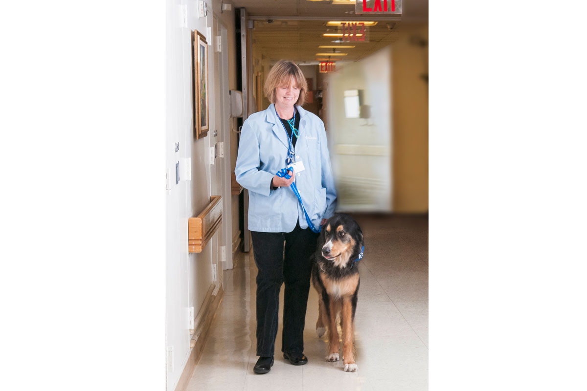
Wagging tails. Wet noses. Soft, warm bellies. It’s not surprising that getting a visit from a therapy dog while receiving cancer treatment boosts the spirits. But these visitors do more than put smiles on the faces of people with cancer — they may actually help some people heal.

“Getting up and moving around after surgery is important to the recovery process, but it’s hard for many people to do,” says Jane Kopelman, who runs the Caring Canines therapy dog program at Memorial Sloan Kettering. Sometimes all it takes is a nudge from a four-legged friend. “I see patients who are willing to get out of bed, and even take a walk down the hallway, just so they can spend more time with one of the dogs,” she says.

Making the Rounds

Over the past 12 years, MSK’s Caring Canines have become a fixture in almost every area of patient care. The program started on a single floor with seven dogs. Now, about 50 therapy dogs and their handlers, who are all volunteers, make the rounds throughout MSK. Both handlers and pups sport identification badges so that passersby know that they are the real deal.

In addition to visiting inpatients, therapy dogs also make regular visits to MSK’s outpatient locations, including its regional sites. When MSK’s newest facility, the David H. Koch Center for Cancer Care at Memorial Sloan Kettering Cancer Center, opened to patients in January 2020, the Caring Canines were there.

Jane Kopelman and her dog Wally
Jane Kopelman and her dog, Wally, visit Memorial Hospital in 2010.

Ms. Kopelman and her mixed-breed dog, Wally, were one of the first volunteer pairs to come to MSK when the Caring Canines program launched in 2007. She quickly realized she wanted to get more involved, and she officially became the Caring Canines consultant in 2014. “It was clear that the program was going to grow a lot, and they needed someone to safely run it,” she says.

Safety is important, especially because many people being treated for cancer have compromised immune systems. Therapy dogs need a host of vaccines and frequent health screenings. They also get bathed and groomed right before visiting MSK. To make sure they don’t tire out, there are limits on how many hours the dogs can work in each shift.

Furry Motivators

In addition to their work as cheer bringers, some of MSK’s Caring Canines have taken part in published research. This got them one step closer to earning their white lab coats — and maybe eventually, their PhD(og)s.

In April 2018, a team led by Pamela Ginex, who was then a nurse researcher at MSK, published a study in the Clinical Journal of Oncology Nursing (CJON) that looked at the benefits of therapy dogs for people recovering from surgery on the 15th floor of Memorial Hospital. At the time, Caring Canines were not yet visiting this floor. People who’d had surgery, as well as their family members and the nursing staff, completed questionnaires on their levels of stress and sense of well-being before and after the visits. This was the first-ever published study to also look at whether therapy dogs can improve job satisfaction for hospital staff in an inpatient setting.

“We had many patients who were having a hard time getting out of bed after having abdominal surgery,” says MSK nurse practitioner Mary Montefusco, who was a co-author on the CJON study. “But when they heard the dogs were on the way, they wanted to get up and sit in a chair so it would be easier to pet them.”

The improvement seen in people who received visits from the dogs was not statistically significant when compared with those who didn’t get visits. (Studies done at other hospitals have shown stronger gains.) The MSK researchers believe this was the case because people progressively feel better as they recover from surgery — whether they get visits from therapy dogs or not.

Anecdotally, however, the sessions were a huge success, not only for the people with cancer but also for their family members and even for staff. “It always brightens my day when I see one of the dogs,” Ms. Montefusco says.

Since the research was published, the Caring Canines have continued visiting the 15th floor and have expanded into more inpatient areas of the hospital. Most recently they’ve started working on the 18th floor, where people recover from surgery for lung cancer. People who are receiving blood and marrow stem cell transplants can now have pooch meet-and-greets, if their immune systems are strong enough. The dogs have started visiting MSK Kids patients as well, meeting up in a play area rather than patients’ rooms to avoid disruptions.

It’s a “Ruff” Job

It takes a rare breed to make it through the rigorous training process that qualifies a dog to work in a medical setting. Not literally, though. Ms. Kopelman says that any dog is capable of becoming a therapy dog, from the tiniest Chihuahua to some really big woofers. The traits required are more about an individual dog’s personality and temperament. “You need a dog that can roll with anything,” she says. “You don’t want them to get flustered and start barking.”

Their handlers also need to complete rigorous training. They are sometimes brought into situations that are emotionally difficult, such as visiting people who are receiving intensive care or those who are at the end of life. “The dog handlers are a vital part of the team,” Ms. Kopelman says.

“Currently, the demand for therapy dogs is much higher than the supply,” Ms. Kopelman concludes. “We would love to have more dogs and volunteers. The most common thing we hear from patients when we arrive at their bedside with a dog is ‘I’ve been waiting all day for this visit.’ ”

A Changing Melanoma Landscape: How Research Has Improved the Outlook for People with Advanced Disease

Source: Memorial Sloan Kettering - On Cancer
Date: 05/04/2020
Link to original
Image of article

In recent years, advanced melanoma has been transformed from a disease that was almost always fatal to one that often can be brought under control for years or even cured. Thanks to new drugs, people with advanced disease now have a five-year survival rate of about 50%.

“The landscape for melanoma has changed dramatically,” says Memorial Sloan Kettering medical oncologist Paul Chapman, who specializes in treating advanced forms of the disease. “Here at Memorial, because we were involved in the early trials, outcomes started to really improve in 2006 or 2007. For the rest of the world, they changed in the early 2010s, after these new drugs were approved and went into wider use.”

Treatments for melanoma have advanced on two fronts: immunotherapy and targeted therapy — both of which have contributed to the remarkable changes that have occurred. But some patients don’t do as well with those drugs, so researchers continue to focus on new therapies and treatment combinations.

Harnessing the Immune System to Fight Cancer

In the area of immunotherapy, the first drug to show significant promise was ipilimumab (Yervoy®). The drug works by exploiting the ability of the body’s own immune system to attack cancer. Specifically, it blocks the activity of a protein called CTLA-4. This takes the brakes off the immune system and enables immune cells called T cells to go after cancer.

Ipilimumab was developed in 1996 by immunologist James Allison, who served as Chair of the Sloan Kettering Institute Immunology Program between 2004 and 2012. Dr. Allison later won a Nobel Prize for this work. MSK physician-scientist Jedd Wolchok led the clinical trials that resulted in the drug’s approval by the US Food and Drug Administration in 2011.

Additional immunotherapy drugs for melanoma, including pembrolizumab (Keytruda®) and nivolumab (Opdivo®), soon followed. They work in a similar way but block a different protein, called PD-1. (Still more immunotherapy drugs that block a related protein called PD-L1 have also been approved.)

Research led by Dr. Wolchok and presented in 2015 showed that, for many people, the combination of ipilimumab and nivolumab was safe and worked better than either drug on its own. That therapy is now standard for many people with metastatic melanoma.

Since the approval of these immunotherapy drugs for melanoma, they have also become a standard treatment for several other types of cancer, including lung cancerbladder cancer, and head and neck cancers. Investigators are continuing to study how these drugs work in order to optimize their use and extend these treatments to more people with cancer.

Targeting the Factors that Drive Cancer

The first-ever targeted drug for melanoma to show a profound effect was vemurafenib (Zelboraf®). It targets a specific mutation in a gene called BRAF and blocks its cancer-causing actions. The mutation is found in about half of all melanomas.

“The drug was designed specifically for people whose cancer contained this mutation,” says Dr. Chapman, who led the phase III trial that resulted in the FDA’s approval of the drug in 2011. “When we saw how well vemurafenib was working, it was a very exciting time.”

Since it was approved for melanoma, vemurafenib has also been approved for cases of a rare blood disorder called Erdheim-Chester disease that have the same BRAF mutation. Other drugs that block the BRAF protein have also been approved.

Another class of targeted therapy for melanoma is called a MEK inhibitor. These drugs block the activity of a growth pathway that is often overactive in melanoma and other cancers. They may be used alone or in combination with BRAF inhibitors.

“Unfortunately, the data show that about 80% of people will eventually develop resistance to BRAF and MEK inhibitors,” Dr. Chapman says. “We’re now looking at new strategies for delivering these drugs.”

Research Aims at Further Gains

“It turns out that melanoma is one of the most responsive cancers to both immunotherapy and targeted therapy,” Dr. Chapman says. “The challenge is to learn more about how to treat the 50% of people whose tumors don’t respond to these treatments.”

Research from MSK and other institutions published in 2013 found that the combination of vemurafenib and ipilimumab led to significant side effects, and therefore the drugs should not be used together. But since then, investigators at MSK and elsewhere have been studying new combinations with different kinds of drugs.

“We’re trying to figure out what’s different about these tumors and if there is some way that we can convert a nonresponsive tumor to a responsive one,” Dr. Chapman adds. “One strategy involves introducing inflammatory molecules to the tumor to see if we can convert it to an environment that’s more receptive to T cell activity.”

Another important aspect of current research is to figure out the smallest dose that can be given to people with cancer while still achieving beneficial effects. “Toxicity and side effects are always a concern when treating any kind of cancer,” Dr. Chapman says. “Part of the history of oncology, whether you’re talking about chemotherapy or some of these more recent drugs, is looking at how much you can dial a treatment back and still cure people. This is an important area of research going forward.”