Crowdsourcing Science: Using Competition to Drive Creativity

Source: National Institute of General Medical Sciences - Biomedical Beat Blog
Date: 02/05/2020
Link to original
Image of article

Historically, crowdsourcing has played an important role in certain fields of scientific research. Wildlife biologists often rely on members of the public to monitor animal populations. Using backyard telescopes, amateur astronomers provide images and measurements that lead to important discoveries about the universe. And many meteorologists use data collected by citizen scientists to study weather conditions and patterns.

Now, thanks largely to advances in computing, researchers in computational biology and data science are harnessing the power of the masses and making discoveries that provide valuable insights into human health.

Tackling Data on Complex Diseases 

Trey Ideker, Ph.D. , a professor of medicine at the University of California, San Diego (UCSD), has used crowdsourcing in his graduate-level bioinformatics classes to analyze results from genome-wide association studies. These studies allow researchers to identify particular gene variations that are linked with a disease or another trait.

Some diseases are triggered by single gene mutations or changes, but most conditions are much more complex. A combination of gene variations as well as environmental and other factors influence disease development.

“Common diseases like diabetes, cancer, heart disease, and neurological and psychiatric disorders have hundreds or even thousands of genes that are contributing to them,” Dr. Ideker says. “When you have diseases that involve so many genes, and the massive amount of data that’s associated with those genes, it’s hard to find connections.”

Students in Dr. Ideker’s class applied a classroom approach to crowdsourcing for a project on schizophrenia. The challenge was to develop computer algorithms that could generate a ranked list of 100 genes associated with schizophrenia. Students were given data on gene variants from more than 51,000 individuals.

“Setting this up as a competition pushed the students to explore different methods,” says UCSD graduate student Samson Fong, the teaching assistant for the class. “In a regular lab setting, you might have one or two people working on a problem. In this case, we allowed the students to develop eight or nine different approaches, which we could compare side by side. We learned much more about which methods worked and which didn’t.”

Dr. Ideker adds that running a competition in a classroom setting, rather than within the scientific community, encouraged collaboration as well as competition.

Fong agrees, noting that students learned a lot from each other. Additionally, they were guided to take their projects in different directions, to ensure a wider range of solutions. He explains that because students worked on teams instead of individually, they tackled much more complex problems than they could have on their own.

Advancing Science with a Combination of Computational Methods

The winning method from the competition led to a computational approach, called Network-Assisted Genomic Association, published in iScience. This approach outperformed other methods in identifying known disease genes and in how well the results from the analysis could be replicated. Dr. Ideker has already used the competition framework in another class to develop algorithms related to computational challenges in structural biology.

“This is a field that’s evolving so quickly that for most problems, there is unlikely to be only one computational method that will work,” Dr. Ideker says. “With this classroom approach, we see how the research can benefit from sampling a bouquet of different ideas. You’re pushing science forward in a really nice way.”

Dr. Ideker’s work is supported by NIGMS grant P41GM103504.
How Errors in Divvying Up Chromosomes Lead to Defects in Cells

Source: National Institutes of General Medical Sciences - Biomedical Beat Blog
Date: 03/25/2020
Link to original
Image of article

Mitosis is fundamental among all organisms for reproduction, growth, and cell replacement. When a cell divides, it’s vital that the two new daughter cells maintain the same genes as the parent.

In one step of mitosis, chromosomes are segregated into two groups, which will go into the two new daughter cells. But if the chromosomes don’t divide properly, one daughter cell may have too many and the other too few. Having the wrong number of chromosomes, a condition called aneuploidy, can trigger cells to grow out of control.

How chromosome segregation errors disrupt cell division is an important area of research. Although it’s been studied for decades, new aspects are still being uncovered and much remains unknown. NIGMS-funded scientists are studying different aspects of mitosis and chromosome segregation. Understanding the details can provide vital insight into an essential biological process and may also be the key to developing better drugs for cancer and other diseases.

Decoding the Mechanics of Chromosome Segregation

Sophie Dumont, Ph.D.  of the University of California, San Francisco, originally trained in physics, but fell in love with biology during graduate school. Her background in physics continues to inform her work, which focuses largely on the mechanical forces that help cells organize themselves during mitosis.

In particular, she’s studying the mitotic spindle (the structure of microtubule filaments that pulls the chromosome pairs apart) and the kinetochore (the protein structure that attaches the chromosome to the spindle).The mitotic spindle and kinetochores are essential to cell organization during mitosis. Credit: Judith Stoffer and NIGMS.

“We’re trying to figure out how these structures, which are made of many small parts, robustly generate and respond to the force that’s needed to move the chromosomes over large distances in the cell,” she explains.

Dr. Dumont’s lab works with human cells, but she also uses a surprising model—kidney cells from rat kangaroos, a small marsupial. Rat kangaroo cells can be helpful because they have many fewer chromosomes than human cells, which makes it easier to follow a single chromosome’s trajectory during mitosis.

“Over millions of years, evolution has found a lot of clever solutions to the challenges of proper chromosome segregation,” she says. “What’s exciting to me about my research is that not only are we making discoveries that have implications for human health, but we are gaining an understanding of how nature can build a diversity of complex structures with very simple parts.”

Recreating Important Processes in the Lab

Chip Asbury, Ph.D. , a scientist at the University of Washington in Seattle, also studies the mechanics of mitosis and chromosome segregation.

“I think of the mitotic spindle as a kind of machine,” he says. “Its job is to move the chromosomes in the appropriate way, while at the same time detecting any errors that may have occurred.”

Dr. Asbury’s lab conducts experiments with the spindle and related components that have been isolated or reconstructed outside the cell. Because kinetochores are made of hundreds of proteins, isolating individual parts of them allows the lab to do experiments they couldn’t do with whole cells.Two Trypanosoma brucei seen through a microscope. Credit: Jeffrey DeGrasse, Rockefeller University.

One experiment introduces tiny artificial cargoes to the kinetochores, enabling Dr. Asbury to recreate and observe how the microtubules form attachments. In addition, Dr. Asbury has a project focusing on mitosis in trypanosomes, a unicellular organism that causes sleeping sickness. This work is a collaboration with Bungo Akiyoshi, Ph.D. , and it may lead to new drugs for targeting these parasites.

“I’m drawn to studying these kinds of fundamental processes because not only are they fascinating, but all life depends on them,” Dr. Asbury says. “Ultimately, our ability to fight complex diseases is limited by our fundamental lack of understanding about what’s happening inside the cell. Understanding how kinetochores work will eventually enable us to develop smarter drugs.”

Linking Chromosomal Errors with Cancer

Jennifer (Jake) DeLuca, Ph.D. , a researcher at Colorado State University in Fort Collins, is looking at how errors in chromosome segregation can lead to cancer. She aims to determine whether it’s possible to develop cancer drugs that target defective segregation.

“The strength of the attachment of the microtubules at the kinetochore must be very tightly controlled,” she explains. “We know that these attachments are misregulated in cancer cells, so we’re working backwards to find out why that is the case.”A human cell undergoing mitosis with the microtubules shown in green, chromosomes shown in blue, and kinetochores shown in red.

To study the chromosome malfunctions seen in cancer, Dr. DeLuca and her team recreate them in human cell models. They transform the cells with cancer-causing genes and then use a number of imaging techniques to watch mitosis in the cells. This allows them to get at the heart of how different proteins regulate microtubule connections in cancer cells.

One of the long-term goals of this research is to figure out how to target these proteins with drugs. “Although we are a basic research lab, not a cancer lab, our work has opened up a huge toolbox of targets to explore,” Dr. DeLuca concludes. “The integration of basic research with clinical research is critical for developing effective new therapies.”

Dr. Dumont’s research is supported by NIGMS grants DP2GM119177 and R01GM134132. Dr. Asbury’s research is supported by R01GM079373 and P01GM105537. Dr. DeLuca’s research is supported by R35GM130365.

Twisting and Turning: Unraveling What Causes Asymmetry

Source: National Institute of General Medical Sciences - Biomedical Beat Blog
Date: 04/02/2020
Link to original
Image of article

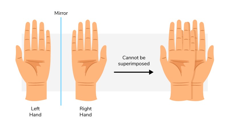
Asymmetry in our bodies plays an important role in how they work, affecting everything from function of internal systems to the placement and shape of organs. Take a look at your hands. They are mirror images of each other, but they’re not identical. No matter how you rotate them or flip them around, they will never be the same. This is an example of chirality, which is a particular type of asymmetry. Something is chiral if it can’t overlap on its mirror image.An image of a pair of hands, palms facing up. An arrow points to another image of the left hand on top of the right, both palms still facing up, illustrating that they can’t be superimposed. Our hands are chiral: They’re mirror images but aren’t identical.

Scientists are exploring the role of chirality and other types of asymmetry in early embryonic development. Understanding this relationship during normal development is important for figuring out how it sometimes goes wrong, leading to birth defects and other medical problems.

Decoding the Causes of Chirality

Michael Ostap , Ph.D., a professor of physiology at the Perelman School of Medicine at the University of Pennsylvania in Philadelphia, is studying how molecules interact to build cell structures that contribute to chirality in living things. His research focuses on the motors in cells, including a motor protein called myosin 1D, which plays an important role in generating chirality.

Dr. Ostap and his lab, along with Stéphane Noselli’s team  in France, examined how myosin 1D triggers chirality during the development of fruit flies. Dr. Noselli’s lab stimulated the production of myosin 1D during the early development of fruit fly organs that usually exhibit symmetry, including the epidermis (outer skin layer) and the trachea (similar to the windpipe). They found that the presence of this protein caused the cells to wind around each other in a spiral shape. The whole fly larva twisted into this spiral, and the spirals were chiral—they always turned in the same direction.

Understanding a Protein’s Push and Pull

Further examination by the Ostap lab revealed that myosin 1D induced spiraling of another protein called actin. Actin proteins form filaments required for cells to move and change shape. In this case, the researchers found that motor activity of the myosin changes the shapes of the cells, so they form tissues in a circular, counterclockwise geometry. How these molecular interactions lead to changes in cell shape, structure, and form remains a fascinating mystery, Dr. Ostap says. Unraveling these mysteries is an important step in developing better ways to treat certain diseases. 

The Ostap lab is continuing to study myosin 1D in flies with a bottom-up approach—from protein to cell to tissue. “We know that this protein is important for chirality,” Dr. Ostap explains. “We’re focused on the biophysical properties of why that’s the case. For example, we are studying myosin 1D’s biochemical and structural properties to try to learn more about how it makes these actin filaments turn.”

Dr. Ostap says studying myosin 1D’s activity in vertebrate research organisms such as mice, chickens, and zebrafish is important as well. A vertebrate’s body has many more components and more complicated interactions than a fruit fly’s body. But some of the same proteins may be important across organisms. “How similar these chiral cues are is not known yet known, and something we plan to study,” he says.

Dr. Ostap’s research is supported in part by NIGMS grant R37GM057247.

Why Salmonella wants its host to have a healthy appetite

Source: Cell Press
Date: 01/26/17
Link to original
Image of article

Anyone who’s ever had the flu or a bad cold can relate to the lethargy, sleepiness, and an increased sensitivity to pain that often result when a pathogen infects a host. A Salk Institute study, published January 26 in Cell, looked at one of the most well-known sickness behaviors–loss of appetite–in mice and found, surprisingly, that when a bacteria reduces its own virulence (how sick it makes the host) by blocking this anorexic response, it actually increases mouse survival and helps the pathogen spread because more food means more infected feces.

“Traditionally in infectious disease, we think that the stronger a pathogen’s ability is to cause disease, the greater its potential is to be transmitted to other hosts,” says senior author Janelle Ayres, an assistant professor in immunobiology and microbial pathogenesis at the Salk Institute for Biological Studies. “But we discovered a pathogen that has evolved to become less dangerous to its host. By employing this strategy, it’s easier for the pathogen to spread to other hosts.”

In the study, the investigators looked at Salmonella Typhimurium, a natural intestinal pathogen in mice (as well as humans) that can easily be transmitted to new hosts. Previous work looking at the connection between Salmonella and loss of appetite has mostly involved injecting a microbe or microbial products directly into the circulation of an animal model and studying its effect, but Ayres’ group infected the animals orally–thus mimicking the bacteria’s route of infection (it spreads from mouse to mouse when the animals eat each other’s contaminated feces).

“Host response is only half of the infectious disease equation. We wanted to understand how the bacteria’s behavior is affected by the host’s loss of appetite, as well,” Ayres says. “What a pathogen wants is a steady supply of nutrients, a stable niche so it can replicate, and a reliable mode of transmission.” In this case, taming the behavior of the pathogen by enabling the mice to take in more nutrition helped keep the mouse healthy, produce more feces, and then spread infection to other animals.

Further investigation revealed the mechanism by which Salmonella Typhimurium inhibits loss of appetite. Sickness behaviors are in large part mediated by a cytokine–a type of molecule involved in cell-to-cell communication–that sends a signal to the hypothalamus, a region of the brain controlling appetite. But this particular Salmonella blocks activation of the cytokine in the intestines, preventing the gut from signaling to the brain.

Ayres says she anticipates finding a similar strategy in other microbes, noting that genes similar to the one known to be important in blocking cytokine activation in SalmonellaTyphimurium also are found in other pathogens. “But a more interesting place to look is at the components of the microbiome, especially the human microbiome,” she notes.

“When an infection in the host affects appetite, the microbiome is also potentially compromised by the loss of nutrition. I expect to find that the microbiome has evolved strategies to block this sickness response,” Ayres adds.

This is something her research group plans to study.

The researchers hope that one day, their findings may lead to a better understanding of infection transmission and new ways to treat infections by supplementing patients with nutrition rather than treating them with antibiotics. The goal would be to give patients a treatment that would also prevent them from spreading their cold or fever to others.

Inactivity in obese mice linked to a decreased motivation to move

Source: Cell Press
Date: 12/29/16
Link to original
Image of article

Starting a regular program at the gym is a common New Year’s resolution, but it’s one that most people are unable to stick with for very long. Now a study done in mice is providing clues about one of the reasons why it may be hard for so many people to stick with an exercise program. The investigators found that in obese mice, physical inactivity results from altered dopamine receptors rather than excess body weight. The report appears in Cell Metabolism on December 29.

“We know that physical activity is linked to overall good health, but not much is known about why people or animals with obesity are less active,” says the study’s senior author Alexxai V. Kravitz, an investigator in the Diabetes, Endocrinology, and Obesity Branch at the National Institute of Diabetes and Digestive and Kidney Diseases–part of the National Institutes of Health. “There’s a common belief that obese animals don’t move as much because carrying extra body weight is physically disabling. But our findings suggest that assumption doesn’t explain the whole story.”

Kravitz has a background in studying Parkinson’s disease, and when he began conducting obesity research a few years ago, he was struck by similarities in behavior between obese mice and Parkinsonian mice. Based on that observation, he hypothesized that the reason the mice were inactive was due to dysfunction in their dopamine systems.

“Other studies have connected dopamine signaling defects to obesity, but most of them have looked at reward processing–how animals feel when they eat different foods,” Kravitz says. “We looked at something simpler: dopamine is critical for movement, and obesity is associated with a lack of movement. Can problems with dopamine signaling alone explain the inactivity?”

In the study, mice were fed either a standard or a high-fat diet for 18 weeks. Beginning in the second week, the mice on the unhealthy diet had higher body weight. By the fourth week, these mice spent less time moving and got around much more slowly when they did move. Surprisingly, the mice on high-fat diet moved less before they gained the majority of the weight, suggesting that the excess weight alone was not responsible for the reduced movements.

The investigators looked at six different components in the dopamine signaling pathway and found that the obese, inactive mice had deficits in the D2 dopamine receptor. “There are probably other factors involved as well, but the deficit in D2 is sufficient to explain the lack of activity,” says Danielle Friend, first author and former NIDDK postdoctoral fellow.

The team also studied the connection between inactivity and weight gain, to determine if it was causative. By studying lean mice that were engineered to have the same defect in the D2 receptor, they found that those mice did not gain weight more readily on a high-fat diet, despite their lack of inactivity, suggesting that weight gain was compounded once the mice start moving less.

“In many cases, willpower is invoked as a way to modify behavior,” Kravitz says. “But if we don’t understand the underlying physical basis for that behavior, it’s difficult to say that willpower alone can solve it.”

He adds that if we begin to decipher the physiological causes for why people with obesity are less active, it may also help reduce some of the stigma that they face. Future research will focus on how unhealthy eating affects dopamine signaling. The researchers also plan to look at how quickly the mice recover to normal activity levels once they begin eating a healthy diet and losing weight.

Transplanted interneurons can help reduce fear in mice

Source: Cell Press
Date: 12/08/16
Link to original
Image of article

The expression “once bitten, twice shy” is an illustration of how a bad experience can induce fear and caution. How to effectively reduce the memory of aversive events is a fundamental question in neuroscience. Scientists in China are reporting that by transplanting mouse embryonic interneurons into the brains of mice and combining that procedure with training to lessen fear, they can help to reduce the fear response. The study is being published December 8 in Neuron.

“Anxiety and fear-related disorders such as post-traumatic stress disorder [PTSD] cause great suffering and impose high costs to society,” says Yong-Chun Yu, a professor at the Institutes of Brain Science at Fudan University in Shanghai and the study’s senior author. “Pharmacological and behavioral treatments of PTSD can reduce symptoms, but many people tend to relapse. There’s a pressing need for new strategies to treat these refractory cases.”

In the study, the researchers used traditional conditioning to instill fear in the mice. They exposed them to a sound as a neutral stimulus, followed by a mild shock to the foot. To determine the level of fear, they measured the amount of time the mice exhibited freezing behavior–the natural sympathetic fear response in prey animals that is indicated by crouching. They then conducted fear extinction training, in which the mice were exposed to the sound but not the shock. After a few rounds, the freezing response times were significantly reduced.

To determine the contribution that transplanting immature interneurons into the amygdala–a brain structure known to be involved in processing of fear and other emotions–could have on fear extinction training, they inserted medial ganglionic eminence (MGE) cells taken from embryos into the amygdala regions of mature mice. The transplanted cells were labeled with green fluorescent protein, enabling the researchers to experimentally confirm that the new cells were integrating into the brains’ circuits.

“We found that although the transplanted interneurons did not alter the formation of fear memories, they reduced recovery and renewal of fear after extinction training,” Yu says. However, transplantation of the neurons alone was not enough to reduce fear memories, indicating that the MGE cells were boosting the effectiveness of that training.

“Unexpectedly, we observed that the erasure of fear memory is facilitated only by transplanted immature interneurons–two weeks after transplantation,” he adds. “Previous studies had indicated that transplanted MGE cells induce plasticity when they are relatively mature–four weeks after transplantation.”

Further studies indicated that the transplanted immature interneurons reactivated a juvenile-like plasticity in the mature amygdala. “Likely related to the changes in the expression of perineuronal nets (PNNs), which are responsible for synaptic stabilization, we found that transplanted immature neurons enhance synaptic plasticity in the amygdala’s circuits by disrupting PNNs, converting the amygdala to a juvenile stage,” Yu says.

Additional experiments are required to determine how these transplanted immature interneurons rejuvenate the mature circuits. “We still don’t know the mechanism by which these immature neurons modulate the fear extinction behavior in the mice,” he concludes. “We also need to determine the exact subtype of transplanted interneurons and the exact subregion in the amygdala that are responsible for these behavioral effects.”

Gut microbe movements regulate host circadian rhythms

Source: Cell Press
Date: 12/01/16
Link to original
Image of article

Even gut microbes have a routine. Like clockwork, they start their day in one part of the intestinal lining, move a few micrometers to the left, maybe the right, and then return to their original position. New research in mice now reveals that the regular timing of these small movements can influence a host animal’s circadian rhythms by exposing gut tissue to different microbes and their metabolites as the day goes by. Disruption of this dance can affect the host. The study appears December 1 in Cell.

“This research highlights how interconnected the behavior is between prokaryotes and eukaryotes, between mammalian organisms and the microbes that live inside them,” says Eran Elinav, an immunologist at the Weizmann Institute of Science, who led the work with co-senior author Eran Segal, a computational biologist also at the Weizmann. “These groups interact with and are affected by each other in a way that can’t be separated.”

The new study had three major findings:

  • The microbiome on the surface layer of the gut undergoes rhythmical changes in its “biogeographical” localization throughout the day and night; thus, the surface cells are exposed to different numbers and different species of bacteria over the course of a day. “This tango between the two partners adds mechanistic insight into this relationship,” Elinav says.
  • The circadian changes of the gut microbiome have profound effects on host physiology, and unexpectedly, they affect tissue that is far away from the gut, such as the liver, whose gene expression changes in tandem with the gut microbiome rhythmicity. “As such,” adds Elinav, “disturbances in the rhythmic microbiome result in impairment in vital diurnal liver functions such as drug metabolism and detoxification.”
  • The circadian rhythm of the host is deeply dependent on the gut microbiota oscillations. Although some circadian machinery in the host was maintained by its own internal clock, other components of the circadian clock had their normal rhythms destroyed. Most surprising, another set of genes in the host that normally exhibit no circadian rhythms stepped in and took over after the microbial rhythms were disrupted.

Previous work by Elinav and Segal revealed that our biological clocks work in tandem with the biological clocks in our microbiota and that disrupting sleep-wake patterns and feeding times in mice induced changes in the microbiome in the gut.

“Circadian rhythms are a way of adapting to changes in light and dark, metabolic changes, and the timing of when we eat,” says Segal. “Other studies have shown the importance of the microbiome in metabolism and its effect on health and disease. Now, we’ve shown for the first time how circadian rhythms in the microbiota have an effect on circadian rhythms in the host.”

The investigators say their work has potential implications for human health in two important ways. First of all, because drugs ranging from acetaminophen to chemotherapy are metabolized in the liver, understanding — and potentially being able to manipulate — the circadian rhythms of our microbiota could affect how and when medications are administered.

Second, understanding more about this relationship could help to eventually intervene in health problems like obesity and metabolic syndrome, which are more common in people whose circadian rhythms are frequently disrupted due to shift work or jet lag.

“What we learned from this study is that there’s a very tight interconnectivity between the microbiome and the host. We should think of it now as one supraorganism that can’t be separated,” Segal says. “We have to fully integrate our thinking with regard to any substance that we consume.”

Tobacco plants engineered to manufacture high yields of malaria drug

Source: Cell Press
Date: 10/20/16
Link to original
Image of article

In 2015, the Nobel Prize in Physiology or Medicine was awarded in part for the discovery of artemisinin, a plant-derived compound that’s proven to be a lifesaver in treating malaria. Yet many people who need the drug are not able to access it, in part because it’s difficult to grow the plant that is the compound’s source. Now, research has shown that tobacco plants can be engineered to manufacture the drug at therapeutic levels. The study appears October 20 in Molecular Plant.

“Artemisinin treats malaria faster than any other drug. It can clear the pathogen from the bloodstream within 48 hours,” says senior author Shashi Kumar, of the International Centre for Genetic Engineering and Biotechnology in New Delhi, India. “Our research is focused on finding a way to make this drug available to more people.”

Malaria infects more than 200 million people every year, according to the World Health Organization, and kills more than 400,000, mostly in Africa and Southeast Asia. The majority of those who live in malaria-stricken areas cannot afford to buy artemisinin. The drug’s high cost is due to the extraction process and largely to the fact that it’s difficult to grow Artemisia annua (sweet wormword), the plant that is the original source of the drug, in climates where malaria is common, such as in India. Advances in synthetic biology have made it possible to produce the drug in yeast, but the manufacturing process is difficult to scale up.

Earlier studies looked at growing the compound in tobacco–a plant that’s relatively easy to genetically manipulate and that grows well in areas where malaria is endemic. But yields of artemisinin from those plants were low.

In the current paper, Kumar’s team reports using a dual-transformation approach to boost the production of artemisinin in the tobacco plants: they first generated plants that contained transgenic chloroplasts, and the same plants were then manipulated again to insert genes into the nuclear genome as well. “We rationalized the expression of biosynthetic pathway’s gene in different compartment that enabled us to reach the maximum yield from the double transgenic plants,” he says.

Extract from the plants was shown to stop the growth progression of pathogen-infected red blood cells in vitro. Whole cells from the plant were also fed to mice infected with Plasmodium berghei, one of the microbes that causes malaria. The plant product greatly reduced the level of the parasite in the blood. In fact, the researchers found, the whole plant material was more effective in attacking the parasite than pure artemisinin, likely because encapsulation inside the plant cells protected the compound from degradation by digestive enzymes.

But Kumar and his colleagues acknowledge that convincing people to eat tobacco plants is likely to be a hard sell. For that reason, he is collaborating with Henry Daniell, a professor of biochemistry at the University of Pennsylvania and one of the study’s coauthors, with a plan to genetically engineer lettuce plants for producing artemisinin. The lettuce containing the drug can then be freeze dried, ground into a powder, and put into capsules for cost-effective delivery.

“Plant and animal science are increasingly coming together,” Kumar says. “In the near future, you will see more drugs produced inside plants will be commercialized to reduce the drug cost.”

Newly discovered gut organism protects mice from bacterial infections

Source: Cell Press
Date: 10/06/16
Link to original
Image of article

While bacteria are often stars of the gut microbiome, emerging research depicts a more complex picture, where microorganisms from different kingdoms of life are actively working together or fighting against one another. In a study published October 6 in Cell, scientists reveal one example: a newly discovered protist that protects its host mice from intestinal bacterial infections.

“This was a serendipitous finding, but an important one,” says senior author Miriam Merad, a Professor of Oncological Sciences and of Medicine at the Icahn School of Medicine at Mount Sinai. “This study shows how vital it is to go beyond bacteria when studying the microbiome.”

The investigators made the discovery when they realized that mice that had been bred at their own facility had a greater number of immune cells in the gut than mice purchased from an outside vendor. Graduate student Aleksey Chudnovskiy, the study’s first author, together with postdoctoral fellow Arthur Mortha, decided to figure out why that was the case. When they performed an intestinal cleanse on the two groups of mice, they were surprised to find that the mice from the Mount Sinai facility had flagellated protozoa living in their guts. DNA sequencing revealed that the microorganism was a new protozoan parasite, which they named Tritrichomonas musculis (T. mu).

Further investigations showed that when this protist was given to the mice that didn’t have it, they, too, had an increase in the number of immune cells in their guts and also increased inflammatory cytokines. The researchers set out to discover the underlying mechanism. They found that T. mu activates the inflammasome in the gut epithelial cells of the mice, which in turn led to the activation of cytokines. They also found that dendritic cells were required to induce inflammation.

To determine whether colonization of T. mu in the gut affected the mice’s ability to fight off infection, they infected mice with Salmonella and found that the animals that had T. mu as part of their microbiome were very resistant to Salmonella infection. “The protective effect of this species is very striking,” Chudnovskiy says.

T. mu was found to be an ortholog of Dientamoeba fragilis, a parasite that’s found in the guts of many humans, but the researchers don’t know if D. fragilis also has a protective effect. It’s something they plan to study. “People from industrialized countries traveling to emerging countries are more susceptible to intestinal infection than the indigenous population,” Merad explains. “It’s possible that protists, which are known to be common in emerging countries, contribute to the protective effect against intestinal pathogenic infections.”

She adds: “The fight against pathogens determined the survival of the human species, and those with stronger immune systems are the ones who survived. It is likely that the microbiome is a big part of the evolutionary process. Thus, identifying those commensals that confer immune strength in exposed communities should help identify novel therapeutics.”

Human stem cells treat spinal cord injury side effects in mice

Source: Cell Press
Date: 10/03/16
Link to original
Image of article

People with spinal cord injuries suffer from many complications in addition to paralysis and numbness. Some of these problems are caused by a lack of the neurotransmitter GABA in the injured spinal cord. Now research in mice is showing that human embryonic stem cells differentiated into medial ganglionic eminence (MGE)-like cells, which produce GABA, may help alleviate two of the most severe side effects–chronic neuropathic pain and bladder dysfunction. The results appear September 22 in Cell Stem Cell.

This study, a collaboration between senior authors Arnold Kriegstein, Director of the Developmental and Stem Cell Biology Program at the University of California, San Francisco (UCSF), and Linda Noble-Haeusslein, a professor in the Departments of Neurological Surgery and Physical Therapy and Rehabilitation Science at UCSF, addressed both neuropathic pain and bladder dysfunction, which are at least in part attributed to overactive spinal cord circuits. “We reasoned if we could take inhibitory neurons and directly place them into the spinal cord in the regions that are overactive, they might integrate into those circuits and suppress the activity,” says Kriegstein.

GABA (gamma-Aminobutyric acid) is an inhibitory neurotransmitter that is found throughout the central nervous system. It plays an important role in reducing the excitability of neurons by binding to receptors that act on synapses. In the study, the investigators used GABAergic inhibitory neuron precursors called MGE-like cells that were derived from human embryonic stem cells.

The neural precursor cells were placed into the spinal cords of mice two weeks after injury had been induced, where they could differentiate into GABA-producing neuron subtypes and form synaptic connections.

“Rather than implanting these cells into the site of injury, at the mid-thoracic level, we injected them in the lumbosacral region, where the circuits are known to be overactive,” says Thomas Fandel, a research specialist at UCSF and the study’s co-first author. “Six months later we could see broad dispersion of the cells in that area. They were integrated into the spinal cord.”

The researchers used several measures to determine whether the stem cells were effective in alleviating neuropathic pain and bladder dysfunction at six months. To assess for bladder control, the mice were placed in cages with filter paper that showed where the mice had urinated. The treated mice had fewer, larger spots, indicating less leakage. Bladder function was also assessed by measuring bladder volume and tension, which confirmed the improved voiding ability of animals receiving transplants.

By six months after transplant, animals exhibited significantly reduced pain sensitivities. Grooming and scratching behaviors were also evaluated, as decreased GABA in the spinal cord can similarly cause pathological itch (pruritus). The researchers found that mice receiving the stem cell transplants showed decreased overgrooming compared to controls.

“The fact that these cells were implanted in the spinal cords two weeks after injury is also important to note,” says Alpa Trivedi, a researcher at UCSF and co-first author of the study. “Many of the current Phase I trials for spinal cord injury are run in the acute phase, which is right after injury. But the vast majority of people with spinal cord injuries are the chronic patient population, and a treatment that might work for them would capture a larger number of patients who are really in need of better treatments.”

Current treatments for neuropathic pain in people with spinal cord injuries most often involve opioids and other pain medications, as well as certain antidepressants, which have many side effects and tend to have limited efficacy. Treatments for bladder dysfunction are often anticholinergics, but these drugs have side effects like dizziness and dry mouth. Botulinum toxin (Botox®) may help with bladder spasms, but the benefits tend to be transient. “The current approaches for treatment are not very effective and clearly more options are needed,” Dr. Fandel says. “Our hope is that this treatment would last a long time, or maybe even be permanent.”

Additional research is needed before the stem cells can be tested in patients, but the researchers have already taken the first step by using human-derived stem cells rather than mouse stem cells in their animal models. “Chronic pain and bladder dysfunction remain significant quality-of-life issues for many people with spinal cord injuries. Inhibitory cell-based neuro-therapy is a new approach and has shown promise to date in early animal studies, warranting further development,” says Cory Nicholas, a co-first author.