Researchers Discover Stem Cells That May Drive Aggressive Behavior in Glioblastoma

Source: Memorial Sloan Kettering - On Cancer
Date: 02/20/2020
Link to original
Image of article

For the past several years, researchers have recognized that the brain tumor glioblastoma is powered by cancer cells called tumor stem cells. Figuring out how tumor stem cells function is important because their ability to survive likely explains why glioblastoma is so hard to treat.

A multicenter team led by scientists at Memorial Sloan Kettering recently reported discovering a likely identity for these tumor stem cells. The leading contenders are called radial glia cells. Radial glia cells play a key role in building fetal brains but were previously thought to disappear after birth. The findings were published January 30 in Stem Cell Reports.

“We can’t say with certainty that radial glia cells are the same as tumor stem cells, but they are now very high on the candidate list,” says physician-scientist Viviane Tabar, Chair of MSK’s Department of Neurosurgery, who was the study’s senior author. “The look and behavior of brain stem cells in the developing brain and the tumor stem cells that we have identified are so similar to each other. This is the first time we’ve seen these features in cells from a human tumor.”

An Unexpected Finding

The first clues about the identity of these tumor stem cells were uncovered by Rong Wang, a research associate in Dr. Tabar’s lab. Dr. Wang was studying tumor tissue that had been removed from patients. She was using the tissue to grow organoid-like structures in petri dishes. Organoids are miniature organs that look and behave very much like their full-size counterparts. They are an increasingly important tool across cancer research for studying tumor development as well as for testing drugs.

Dr. Wang noticed that some cells in the organoids had an unusual shape and exhibited unusual behavior. They had very long processes, or tails, and when they divided, their daughter cells had the same shape. Additionally, when these unusual cells divided, their nuclei jumped over long distances. Dr. Wang recognized that these features are also seen in radial glia.

Further analysis confirmed that these unusual cells also were present in the tissue taken from patients: The team studied samples from dozens of tumors.

“Radial glia cells previously were not thought to persist in adulthood,” Dr. Tabar explains. “If glioblastoma tumors arise from them, that may mean that humans retain some radial glia cells in our brains as adults. The other possibility is that the genetic changes in the cancer turn some of the brain cells back into cells that look very much like radial glia cells.”

A Valuable Collaboration

To learn more about these cells, the Tabar lab collaborated with Dana Pe’er, Chair of the Sloan Kettering Institute’s Computational and Systems Biology Program, as well as with computational biologists at the Wellcome Sanger Institute in the United Kingdom.

They performed studies called single-cell RNA sequencing to look at which genes were expressed, or turned on, in individual cells. The patterns of gene expression observed in the samples taken from patient tumors and those grown in the lab were very similar to what’s seen in radial glia cells from embryos.

“This was a very challenging study to pull together. We took advantage of MSK’s wide range of tumors and access to fresh surgical tissue, as well as the resources of the Computational and Systems Biology Program here,” Dr. Tabar says.

A Potential Explanation for a Cancer’s Aggressive Behavior

Additional research is needed to confirm whether these cells are indeed the same. Another focus of future work will be the role of inflammation. “Inflammation may help bring radial glia-like cancer cells out of dormancy,” Dr. Tabar explains. “This could explain why inflammation can sometimes lead to the worsening of brain tumors.”

Dr. Tabar hopes that the publication of the findings will encourage further analysis and that “as technology advances, there will be easier ways to identify and study these cells,” she concludes.
What’s the Best Way to Screen for Breast Cancer? Abbreviated MRI Beats 3-D Mammography, but Research Is Ongoing

Source: Memorial Sloan Kettering - On Cancer
Date: 02/28/2020
Link to original
Image of article

It’s well understood that women in certain age groups should undergo regular screenings to look for signs of breast cancer. But as technology has progressed, the methods used for screening have evolved.

Memorial Sloan Kettering radiologist Christopher Comstock published a study in the Journal of the American Medical Association (JAMA) on February 25 that shed light on this topic. The trial found that a new test called abbreviated MRI found far more cancers than 3-D mammography in women at average risk who have dense breasts. An abbreviated MRI reduces the length of a standard MRI scan from 45 to 10 minutes.

“About half of all women have dense breasts,” Dr. Comstock notes. Having dense breasts makes it more difficult for cancer to be detected because the dense tissue can obscure cancerous masses.

The most common method for screening women with dense breasts is 3-D mammography. This imaging test creates a three-dimensional view of the breast tissue. It is often combined with ultrasound. Also known as tomosynthesis, 3-D mammography is better than regular 2-D mammography at detecting masses in dense tissue. Abbreviated MRI, which uses fewer images than full MRI, is one of the new approaches being developed that could eventually replace current digital mammography techniques.

The study findings raise concerns that mammograms may not be the best way to screen for cancer among women who are of average risk and have dense breasts.

“Previous research done at MSK and elsewhere has shown that full-breast MRIs are the best imaging method for detecting cancer, but these tests are expensive, time-consuming, and not widely available,” Dr. Comstock explains. “This study was designed to look at the ability of abbreviated MRI to find breast cancer.” The trial also showed women tolerate the MRI with very few side effects and that the centers could perform the test in less than 10 minutes.

A Constantly Evolving Field

When doctors began screening for breast cancer in the 1960s and 1970s, they used standard X-rays. Eventually, they developed more-specialized techniques and equipment for doing mammograms. In the early 2000s, digital mammography, in which images are stored on computers, replaced films. In the past several years, 3-D mammography has replaced 2-D mammography as the standard screening method for women with dense breasts. Digital 2-D mammography is the standard for those who don’t have dense breasts.

More than 1,400 women, ages 40 to 75, participated in the JAMA study. All of them were found to have dense breasts on a prior mammogram, did not have any signs or symptoms of breast cancer, and were of average risk.

The women in the trial were screened with both 3-D mammography and abbreviated breast MRI at 48 centers in the United States and Germany.

In the first year of the study, 23 women were diagnosed with breast cancer. The abbreviated MRI detected 22 out of the 23 breast cancers, while the 3-D mammogram detected only nine out of the 23 cancers. Only one cancer was discovered with 3-D mammography that was not found with abbreviated MRI.

“The findings from this trial are significant,” Dr. Comstock notes. “The abbreviated MRI found about two and a half times more cancers than mammography alone, including ten that were invasive cancers.”

However, he notes that abbreviated MRI is still very new. Many breast-screening centers don’t have MRI machines — especially community hospitals and those in more rural areas. Experts at MSK are figuring out the best way to offer abbreviated MRI, which is not currently covered by insurance, to patients.

The Promise of Other Screening Technologies

Dr. Comstock points to another vascular-based technology that is similar to MRI, called contrast-enhanced digital mammography (CEDM), which he believes shows similar promise for becoming the new standard screening method for women with dense breasts. MSK already offers this test at the Evelyn H. Lauder Breast Centerin Manhattan as well as at all of its regional locations.

“CEDM is a significant improvement over standard 3-D mammography,” says MSK Senior Vice President Larry Norton, a medical oncologist who specializes in treating breast cancer. “One of the advantages of CEDM compared with MRI is that conventional mammography machines can be easily adapted to do CEDM, which means that more centers can offer it.”

“In the near term, CEDM is likely to be much more available and accessible to patients than MRI, and at a lower cost,” Dr. Comstock adds. “It uses a technology that’s already familiar to doctors, which makes it easier to provide.”

Research done at MSK has already shown the benefits of CEDM at detecting cancer in women with dense breasts, compared with conventional 3-D mammography: A study published in Radiology in October 2019 found that the newer technique detected nearly twice as many cancers. These early results suggest that CEDM, which has been offered at MSK since 2012, is a promising way to screen for breast cancer.

Looking Ahead

Dr. Comstock is currently planning a large multicenter trial to assess whether CEDM screening is more accurate in women with dense breasts compared with the combination of 3-D mammography and ultrasound. Called the Contrast-Enhanced Mammography Imaging Screening Trial (CMIST), it is expected to launch within the next few months. CMIST will be conducted at about a dozen centers around the world. “At MSK, we are exploring more-advanced techniques that detect cancers earlier to lead to better treatment,” says Elizabeth Morris, Chief of the Breast Imaging Service at MSK, who is also involved in planning CMIST. “The landscape of breast radiology is ever changing, and here at MSK, we are leading much of this research.”

Deep Understanding of Immunotherapy Helps Patients Cope with Side Effects

Source: Memorial Sloan Kettering - On Cancer
Date: 04/16/2020
Link to original
Image of article

Immunotherapy has changed the outlook for many people with cancer. It offers long-term control or even a cure for tumor types that don’t respond well to other treatments. But because immunotherapy works in a different way than more traditional cancer treatments, such as chemotherapy and radiation, it can lead to new kinds of side effects.

Memorial Sloan Kettering is a leader in developing immunotherapy approaches, including drugs called checkpoint inhibitors. These drugs work by taking the brakes off immune cells and allowing them to go after cancer. But sometimes the immune system becomes too active and attacks healthy tissue. This reaction, called an immune-related adverse event, occurs in about one-third of people taking these drugs. As pioneers in immunotherapy, MSK care providers have rich experience in managing and easing these side effects. This enables most people to complete their cancer treatment and increases the chances that it will ultimately be successful. 

The most common immune-related adverse events caused by checkpoint inhibitors are skin problems, such as rashes, and inflammation of the gastrointestinal tract, which causes problems like diarrhea. Less frequent but potentially serious side effects include inflammation in the heart, liver, kidneys, lungs, and endocrine glands. Overactive immune cells can also affect the joints. This can lead to a painful condition similar to rheumatoid arthritis.

A Leader in Clinical Trials

Some of the earliest clinical trials of checkpoint inhibitor drugs were headed by MSK physician-scientist Jedd Wolchok. From the beginning, Dr. Wolchok and his colleagues — including Alyona Weinstein, a nurse practitioner who works in Dr. Wolchok’s clinic — began seeing immune-system reactions in their patients that looked like autoimmune disorders.

“We’ve known for a long time how to manage the most common side effects from chemotherapy,” such as nausea and reduced blood counts, Ms. Weinstein says. But the side effects from checkpoint inhibitor drugs can be more wide-ranging and unpredictable. “The inflammation caused by an overactive immune system can happen in any part of the body,” she notes. “At MSK, we are careful to screen patients for these side effects early in their treatment so that we can manage them before they become serious.”

A Growing Community of Specialists

As more people have received checkpoint inhibitors, a cadre of experts in immune-related side effects has naturally grown within MSK’s Division of Subspecialty Medicine. Specialists include dermatologists, gastroenterologists, cardiologists, endocrinologists, and more. They focus on health problems other than cancer. But because they work at MSK, they exclusively treat these disorders in people with cancer.

“When patients see the list of potential side effects, they often get very worried,” Ms. Weinstein says. “But in many cases, the appearance of these side effects is an early indication that the drugs are working.” She adds that while some people have no side effects, others may experience more than one serious complication. 

“The management of side effects requires supportive services from many areas beyond medical oncology, and MSK has these specialists,” says Michael Postow, a medical oncologist who specializes in immunotherapy. “Our doctors see these problems a lot, and they’ve developed deep expertise within their areas of specialization.”

Focusing on Health and Quality of Life during and after Treatment

MSK dermatologist Mario Lacouture treats people with skin-related side effects from immunotherapy and other cancer treatments. He recently received a five-year grant from the National Institutes of Health to study the immune-system-related side effects of immunotherapy. The project is a collaboration with National Jewish Health in Denver, a leading center for immunological disorders.

“The big dilemma is that you want to suppress the side effects of immunotherapy enough that patients feel well but not enough that the cancer therapy is no longer active or that patients have additional side effects from immunosuppressive drugs,” Dr. Lacouture explains. “We plan to use the skin as a model to identify what causes these autoimmune reactions so we can develop better ways to treat side effects without reducing the effectiveness of the cancer treatment.”

Other MSK specialists who play a role in treating autoimmune side effects include gastroenterologist David Faleck, cardiologist Dipti Gupta, and endocrinologist Monica Girotra. MSK’s team also closely collaborates with specialists at other area hospitals. This includes rheumatologists at the Hospital for Special Surgery, who are studying arthritis caused by checkpoint inhibitors and treat many of MSK’s patients.

Physical and occupational therapists, as well as specialists in integrative medicine, can help patients cope with pain and mobility problems. Because the gastrointestinal side effects from checkpoint inhibitor drugs can be difficult to treat with medication alone, MSK also has nutritionists who can advise people about the best diets for reducing symptoms related to these complications.

“Our hope is that our patients live a long time, and we know that, unfortunately, autoimmune side effects can continue even after they finish their treatment,” Ms. Weinstein concludes. “We are focused on making sure our patients have a good quality of life in addition to successful treatment for their cancer.”

Research Uncovers Details about How Gut Microbes Influence the Immune System

Source: Memorial Sloan Kettering - On Cancer
Date: 04/22/2020
Link to original
Image of article

The microscopic organisms that live in our intestines perform many jobs to keep us healthy, such as helping our bodies digest nutrients from food. They keep harmful microbes under control and prevent them from taking over the gut, and also play a role in regulating the immune system.

Researchers at Memorial Sloan Kettering are studying the connections between gut microbes and human health. In a study published April 15 in Nature, an MSK team uncovered new findings about an important relationship: the way that gut microbes promote the formation of a type of immune cell called regulatory T cells (Tregs).

Tregs help keep powerful immune responses in check. They are crucial for preventing autoimmunity, when the immune system mistakenly attacks the body. Tregs also play a role in cancer: Cancer is more likely to develop when malfunctioning Tregs lead to chronically inflamed tissue. This is seen in conditions such as inflammatory bowel disease (IBD).

Focusing on the Microbe–T Cell Connection

“Life in all mammals, including humans, is impossible without regulatory T cells,” says the study’s senior author, Alexander Rudensky, Chair of the Immunology Program in the Sloan Kettering Institute and a Howard Hughes Medical Institute investigator. “This study builds on previous research from our lab and others that looked at how these cells are made and why they are so important.

“It’s already been shown that disturbance of the microbial community in the gut is associated with autoimmune and inflammatory diseases, like IBD,” he adds. “These findings have illustrated the importance of the relationship between the microbes that live in our guts and our immune systems.”

To better understand this relationship, Dr. Rudensky and his team study how different microbes in the gut affect the production of Tregs that protect against inflammation and autoimmune conditions. They do this by looking at the molecules that microbes make to carry out their metabolic functions. Then they study how those metabolites in turn influence the manufacturing of Tregs.

In the experiments, the investigators concentrated on a class of metabolites called secondary bile acids. These are produced by gut microbes. Bile acids help the digestion of dietary fats.

Members of Dr. Rudensky’s team focused on a type of gut bacteria belonging to a group called Bacteroides and engineered them to produce a particular bile acid, called isoDCA. They exposed naive T cells, which had not yet developed into a specific T cell type, to isoDCA during the process of their activation. They found that isoDCA caused the immature T cells to become Tregs.

Ultimately, these findings could lead to a bacterial-based therapy for the treatment of IBD associated with imbalanced microbes and diminished Treg activity. (Currently, IBD is treated with anti-inflammatory drugs, which can have many side effects.) But Dr. Rudensky says much more work is needed before this kind of treatment could be tested. Many parts of the process are still not well understood. In addition, researchers would have to do extensive testing in mice before designing a clinical trial. “We plan to continue exploring the possibility of using metabolic products from microbes for the treatment of inflammatory disorders,” he says.

The two first authors on the paper were research fellow Clarissa Campbell and research associate Peter McKenney in Dr. Rudensky’s lab. Investigators in MSK’s Donald B. and Catherine C. Marron Cancer Metabolism Center, including the center’s director, Justin Cross, also contributed to the research.

Novel Tool Enables Study of Rare Acute Myeloid Leukemia Stem Cells

Source: Memorial Sloan Kettering - On Cancer
Date: 04/27/2020
Link to original
Image of article

If you think of cells as factories for making proteins, and DNA as the instructions contained within those factories, RNA is the workforce that actually carries out the manufacturing. Understanding how RNA does its job is essential for figuring out what goes wrong in many diseases, including cancer.

To take the analogy one step further, RNA-binding proteins (RBPs) are tools that RNA uses in the production process. There are more than 1,500 RBPs in any given cell, which creates a challenge for scientists who want to study them on an individual basis. But researchers are looking for ways to overcome this hurdle because RBPs are an important target for the development of new drugs.

In a paper published April 24 in Nature CommunicationsSloan Kettering Institute cancer biologist Michael Kharas, members of his laboratory, and collaborators in the lab of computational biologist Christina Leslie describe a new tool for studying RBPs. In addition to having broad applications for a range of cell types, the team reports that this tool has already uncovered details about one particular RBP, called Musashi-2. Musashi-2 helps stem cells in the blood become more-specialized cell types. It is known to be overly active in acute myeloid leukemia (AML) cells.

“This is an exciting study because it changes how we study RBPs,” Dr. Kharas says. “It also changes what we know about how they function in specific cells.”

Translating a Lab Technique from Flies to Mammals

The experimental technique used in the study is called HyperTRIBE. It was originally developed to study nerve cells from fruit flies. Dr. Kharas says this is the first published study demonstrating that HyperTRIBE can be used in mammalian cells. The cells they used were blood stem cells from mice and leukemia stem cells from mice and humans.

HyperTRIBE uses a technology that is different from current methods for studying RBPs. Other approaches require millions of cells. The biggest benefit of HyperTRIBE is that it works in rare cells that are available only in very small numbers.

“Our study shows that this technique can be used to study RBPs, not just in fruit fly cells but more broadly,” says Dr. Kharas, a member of SKI’s Molecular Pharmacology Program. “This will have global impact for anyone studying RBPs in rare cell populations, whether those are blood stem cells, neurons, germ cells, or other kinds of stem cells.”

New Clues about a Protein’s Role in Leukemia

In the Nature Communications paper, the investigators report that HyperTRIBE has already revealed important findings about Musashi-2 and how it contributes to AML. Dr. Kharas and the other researchers are developing drugs to treat AML that work by blocking Musashi-2, but they still have a lot to learn about how these drugs modify the function of RBPs.

Using this novel tool, Dr. Kharas’s lab learned that Musashi-2 behaves differently in leukemia cells than it does in regular blood stem cells. “We knew that leukemia cells seemed to be more addicted to Musashi-2 for their growth than normal cells,” Dr. Kharas says. “Now we know that’s because Musashi-2 increases its RNA-binding activity and changes how RNA gets translated into proteins in cancer cells compared to normal cells.”

The investigators plan to continue studying why this is the case. Dr. Kharas says it could aid the development of drugs that slow leukemia growth by affecting Musashi-2’s activity while avoiding side effects that could result if Musashi-2 changes the production of healthy cells. “Because HyperTRIBE doesn’t require a large number of cells, we’ll be able to do more experiments to test potential drugs under many different conditions,” he concludes.

Top Dogs: Meet MSK’s Four-Legged Volunteers

Source: Memorial Sloan Kettering - On Cancer
Date: 04/30/2020
Link to original
Image of article

Wagging tails. Wet noses. Soft, warm bellies. It’s not surprising that getting a visit from a therapy dog while receiving cancer treatment boosts the spirits. But these visitors do more than put smiles on the faces of people with cancer — they may actually help some people heal.

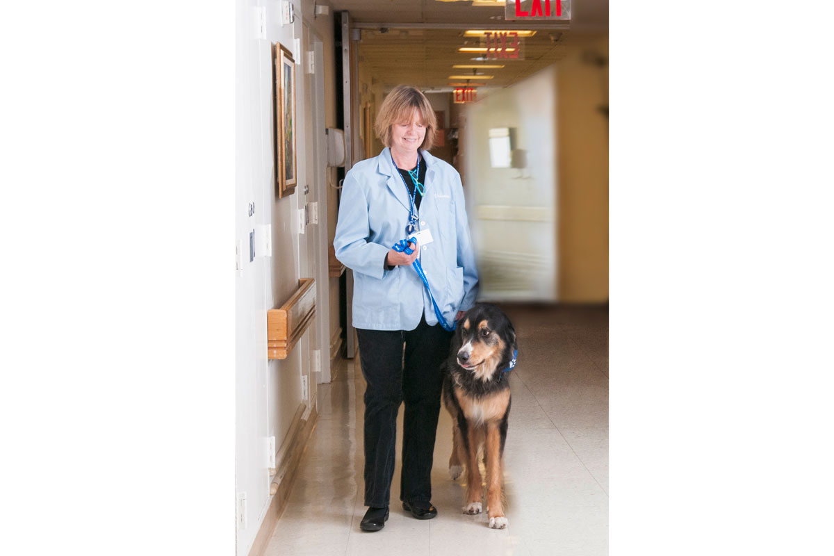
“Getting up and moving around after surgery is important to the recovery process, but it’s hard for many people to do,” says Jane Kopelman, who runs the Caring Canines therapy dog program at Memorial Sloan Kettering. Sometimes all it takes is a nudge from a four-legged friend. “I see patients who are willing to get out of bed, and even take a walk down the hallway, just so they can spend more time with one of the dogs,” she says.

Making the Rounds

Over the past 12 years, MSK’s Caring Canines have become a fixture in almost every area of patient care. The program started on a single floor with seven dogs. Now, about 50 therapy dogs and their handlers, who are all volunteers, make the rounds throughout MSK. Both handlers and pups sport identification badges so that passersby know that they are the real deal.

In addition to visiting inpatients, therapy dogs also make regular visits to MSK’s outpatient locations, including its regional sites. When MSK’s newest facility, the David H. Koch Center for Cancer Care at Memorial Sloan Kettering Cancer Center, opened to patients in January 2020, the Caring Canines were there.

Jane Kopelman and her dog Wally
Jane Kopelman and her dog, Wally, visit Memorial Hospital in 2010.

Ms. Kopelman and her mixed-breed dog, Wally, were one of the first volunteer pairs to come to MSK when the Caring Canines program launched in 2007. She quickly realized she wanted to get more involved, and she officially became the Caring Canines consultant in 2014. “It was clear that the program was going to grow a lot, and they needed someone to safely run it,” she says.

Safety is important, especially because many people being treated for cancer have compromised immune systems. Therapy dogs need a host of vaccines and frequent health screenings. They also get bathed and groomed right before visiting MSK. To make sure they don’t tire out, there are limits on how many hours the dogs can work in each shift.

Furry Motivators

In addition to their work as cheer bringers, some of MSK’s Caring Canines have taken part in published research. This got them one step closer to earning their white lab coats — and maybe eventually, their PhD(og)s.

In April 2018, a team led by Pamela Ginex, who was then a nurse researcher at MSK, published a study in the Clinical Journal of Oncology Nursing (CJON) that looked at the benefits of therapy dogs for people recovering from surgery on the 15th floor of Memorial Hospital. At the time, Caring Canines were not yet visiting this floor. People who’d had surgery, as well as their family members and the nursing staff, completed questionnaires on their levels of stress and sense of well-being before and after the visits. This was the first-ever published study to also look at whether therapy dogs can improve job satisfaction for hospital staff in an inpatient setting.

“We had many patients who were having a hard time getting out of bed after having abdominal surgery,” says MSK nurse practitioner Mary Montefusco, who was a co-author on the CJON study. “But when they heard the dogs were on the way, they wanted to get up and sit in a chair so it would be easier to pet them.”

The improvement seen in people who received visits from the dogs was not statistically significant when compared with those who didn’t get visits. (Studies done at other hospitals have shown stronger gains.) The MSK researchers believe this was the case because people progressively feel better as they recover from surgery — whether they get visits from therapy dogs or not.

Anecdotally, however, the sessions were a huge success, not only for the people with cancer but also for their family members and even for staff. “It always brightens my day when I see one of the dogs,” Ms. Montefusco says.

Since the research was published, the Caring Canines have continued visiting the 15th floor and have expanded into more inpatient areas of the hospital. Most recently they’ve started working on the 18th floor, where people recover from surgery for lung cancer. People who are receiving blood and marrow stem cell transplants can now have pooch meet-and-greets, if their immune systems are strong enough. The dogs have started visiting MSK Kids patients as well, meeting up in a play area rather than patients’ rooms to avoid disruptions.

It’s a “Ruff” Job

It takes a rare breed to make it through the rigorous training process that qualifies a dog to work in a medical setting. Not literally, though. Ms. Kopelman says that any dog is capable of becoming a therapy dog, from the tiniest Chihuahua to some really big woofers. The traits required are more about an individual dog’s personality and temperament. “You need a dog that can roll with anything,” she says. “You don’t want them to get flustered and start barking.”

Their handlers also need to complete rigorous training. They are sometimes brought into situations that are emotionally difficult, such as visiting people who are receiving intensive care or those who are at the end of life. “The dog handlers are a vital part of the team,” Ms. Kopelman says.

“Currently, the demand for therapy dogs is much higher than the supply,” Ms. Kopelman concludes. “We would love to have more dogs and volunteers. The most common thing we hear from patients when we arrive at their bedside with a dog is ‘I’ve been waiting all day for this visit.’ ”

Food Safety during COVID-19: What People with Cancer Should Know

Source: Memorial Sloan Kettering
Date: 04/21/2020
Link to original
Image of article

During the COVID-19 pandemic, we’ve been told to wash our hands frequently, sanitize surfaces, and not touch our faces. On top of that, conflicting reports are circulating online about how to safely handle the food that we’re bringing into our homes.

The challenges of safely buying groceries and getting takeout may be even greater for people with cancer and cancer survivors, who often have weakened immune systems. This makes it harder for their bodies to fight off infections, including COVID-19. It’s important that people with cancer closely follow steps to protect themselves while still getting proper nutrition.

Memorial Sloan Kettering clinical dietitian Cara Anselmo offers guidance for people with cancer and their families on how to carefully get and prepare food during the COVID-19 pandemic.

What safety measures should you take while grocery shopping?

If you are in active treatment for cancer or you have a weakened immune system because of a cancer history or other medical conditions, it’s better to have someone else in your household or a neighbor or friend go to the store for you, or use a delivery service. That’s the most important way you can avoid exposure to COVID-19 or other infectious germs.

But if you do have to go to the store, here are some important tips:

  • Take a grocery list. Before you go, figure out exactly what you need so you can get in and get out fast. This also helps minimize the number of shopping trips.
  • Bring sanitizing wipes to disinfect the handle of the shopping cart or basket.
  • Don’t browse with your hands. When you are at the store, avoid touching food or other products that you don’t intend to buy.
  • Cover your face with a scarf or cloth mask. (In early April, the Centers for Disease Control and Prevention recommended that all Americans wear a face covering to protect themselves from COVID-19.) If you need to adjust your mask, be careful not to touch the part that touches your face.
  • Check to see if your store has special hours for people who are immunocompromised and read the store’s policies on how many shoppers are allowed inside at once. While you are in the store, be careful to maintain a proper distance of six feet from others.

Do you need to disinfect your groceries after bringing them into your home?

At this time, experts are not aware of COVID-19 being spread by food or food packaging. But it’s still important to carefully wash your hands before and after grocery shopping. You should also wash your hands after putting your groceries away.

If you are concerned about the surfaces of packages, you can use disinfectant products to wipe them off. Fresh fruits and vegetables should be carefully washed with running water. Never use Lysol or bleach on fruits and vegetables, since the chemicals could be ingested.

How can you safely get takeout or food delivery from restaurants?

A lot of the advice for grocery shopping is the same for getting takeout or delivery: You should wash your hands before and after touching bags and food packages from restaurants, and before eating.

If you are picking up takeout, make sure you maintain safe distances. If you are getting delivery, pay in advance and ask the delivery person to leave the food outside your door to minimize person-to-person contact.

With either takeout or delivery, you should always transfer food to your own plates and dishes.

People whose immune systems are compromised by cancer or cancer treatment should be careful about eating certain foods from restaurants. This includes things like prepared salads, cut fruit, and deli meats. This is important to avoid food poisoning and not specifically because of COVID-19.

Do you have tips for healthy eating during the COVID-19 pandemic?

If you can’t get fresh produce, frozen or canned fruits and vegetables can be just as healthy. If you normally buy organic foods and you are not able to get them, don’t worry. Organic foods are not necessarily healthier or safer than conventional foods.

In this time of stress and uncertainty, it’s especially important to practice mindful eating. That means you should only eat when you’re hungry. Don’t eat so much that you feel too full. Do the best you can to include plenty of fruits and vegetables, but give yourself permission to eat what tastes good and feels good in your body. There’s no such thing as a perfect diet.

Some dietary supplements are claiming that they improve your immune function and offer protection against COVID-19. What do we know about these products?

Some of these products may be dangerous and potentially even life-threatening. The government has issued warning letters to many companies making these claims about their products.

There have been claims that some familiar supplements, including vitamin C and zinc, may “boost” your immune system. Some studies show that these supplements stimulate the immune system, but the evidence is inconclusive. They can also lead to unwanted effects.

Make sure you talk to your doctor, nurse, or registered dietitian before taking any supplements. Some of these products may also interfere with cancer treatment.

The best way to keep your immune system strong is to eat a balanced diet, limit alcohol, stay hydrated, manage stress, stay as physically active as you can, and get enough sleep.

During this difficult time, many people may be struggling to put food on the table. What are some good resources for MSK patients and their families?

MSK’s Food to Overcome Outcome Disparities program connects patients with a variety of emergency food resources. This includes a food pantry that MSK operates for patients and their families. If you would like more information, you can discuss it with your MSK care team or call MSK’s nutrition office at 212-639-7312.

New York City is offering three free meals per day for both children and adults, with pickup at more than 400 sites. People who are age 60 and older and need assistance can also get home-delivered meals

A Changing Melanoma Landscape: How Research Has Improved the Outlook for People with Advanced Disease

Source: Memorial Sloan Kettering - On Cancer
Date: 05/04/2020
Link to original
Image of article

In recent years, advanced melanoma has been transformed from a disease that was almost always fatal to one that often can be brought under control for years or even cured. Thanks to new drugs, people with advanced disease now have a five-year survival rate of about 50%.

“The landscape for melanoma has changed dramatically,” says Memorial Sloan Kettering medical oncologist Paul Chapman, who specializes in treating advanced forms of the disease. “Here at Memorial, because we were involved in the early trials, outcomes started to really improve in 2006 or 2007. For the rest of the world, they changed in the early 2010s, after these new drugs were approved and went into wider use.”

Treatments for melanoma have advanced on two fronts: immunotherapy and targeted therapy — both of which have contributed to the remarkable changes that have occurred. But some patients don’t do as well with those drugs, so researchers continue to focus on new therapies and treatment combinations.

Harnessing the Immune System to Fight Cancer

In the area of immunotherapy, the first drug to show significant promise was ipilimumab (Yervoy®). The drug works by exploiting the ability of the body’s own immune system to attack cancer. Specifically, it blocks the activity of a protein called CTLA-4. This takes the brakes off the immune system and enables immune cells called T cells to go after cancer.

Ipilimumab was developed in 1996 by immunologist James Allison, who served as Chair of the Sloan Kettering Institute Immunology Program between 2004 and 2012. Dr. Allison later won a Nobel Prize for this work. MSK physician-scientist Jedd Wolchok led the clinical trials that resulted in the drug’s approval by the US Food and Drug Administration in 2011.

Additional immunotherapy drugs for melanoma, including pembrolizumab (Keytruda®) and nivolumab (Opdivo®), soon followed. They work in a similar way but block a different protein, called PD-1. (Still more immunotherapy drugs that block a related protein called PD-L1 have also been approved.)

Research led by Dr. Wolchok and presented in 2015 showed that, for many people, the combination of ipilimumab and nivolumab was safe and worked better than either drug on its own. That therapy is now standard for many people with metastatic melanoma.

Since the approval of these immunotherapy drugs for melanoma, they have also become a standard treatment for several other types of cancer, including lung cancerbladder cancer, and head and neck cancers. Investigators are continuing to study how these drugs work in order to optimize their use and extend these treatments to more people with cancer.

Targeting the Factors that Drive Cancer

The first-ever targeted drug for melanoma to show a profound effect was vemurafenib (Zelboraf®). It targets a specific mutation in a gene called BRAF and blocks its cancer-causing actions. The mutation is found in about half of all melanomas.

“The drug was designed specifically for people whose cancer contained this mutation,” says Dr. Chapman, who led the phase III trial that resulted in the FDA’s approval of the drug in 2011. “When we saw how well vemurafenib was working, it was a very exciting time.”

Since it was approved for melanoma, vemurafenib has also been approved for cases of a rare blood disorder called Erdheim-Chester disease that have the same BRAF mutation. Other drugs that block the BRAF protein have also been approved.

Another class of targeted therapy for melanoma is called a MEK inhibitor. These drugs block the activity of a growth pathway that is often overactive in melanoma and other cancers. They may be used alone or in combination with BRAF inhibitors.

“Unfortunately, the data show that about 80% of people will eventually develop resistance to BRAF and MEK inhibitors,” Dr. Chapman says. “We’re now looking at new strategies for delivering these drugs.”

Research Aims at Further Gains

“It turns out that melanoma is one of the most responsive cancers to both immunotherapy and targeted therapy,” Dr. Chapman says. “The challenge is to learn more about how to treat the 50% of people whose tumors don’t respond to these treatments.”

Research from MSK and other institutions published in 2013 found that the combination of vemurafenib and ipilimumab led to significant side effects, and therefore the drugs should not be used together. But since then, investigators at MSK and elsewhere have been studying new combinations with different kinds of drugs.

“We’re trying to figure out what’s different about these tumors and if there is some way that we can convert a nonresponsive tumor to a responsive one,” Dr. Chapman adds. “One strategy involves introducing inflammatory molecules to the tumor to see if we can convert it to an environment that’s more receptive to T cell activity.”

Another important aspect of current research is to figure out the smallest dose that can be given to people with cancer while still achieving beneficial effects. “Toxicity and side effects are always a concern when treating any kind of cancer,” Dr. Chapman says. “Part of the history of oncology, whether you’re talking about chemotherapy or some of these more recent drugs, is looking at how much you can dial a treatment back and still cure people. This is an important area of research going forward.”

How to Cope with Stress and Anxiety during COVID-19

Source: Memorial Sloan Kettering
Date: 05/08/2020
Link to original
Image of article

The COVID-19 pandemic is taking an emotional toll on people all over the world. Feelings of worry may seem overwhelming, especially if you were already coping with the stress and anxiety of a cancer diagnosis or cancer treatment. 

As a psycho-oncologist, Memorial Sloan Kettering’s Christian Nelson is dedicated to safeguarding the mental health and well-being of people with cancer both during and after treatment. “Feelings of uncertainty and a loss of control are some of the biggest sources of anxiety for people with cancer,” he says. “When you add COVID-19 into the mix, it makes the loss of control feel that much stronger.”

Dr. Nelson, who is Chief of the Psychiatry Service in the Department of Psychiatry and Behavioral Sciences, adds, “COVID-19 causes such intense feelings of uncertainty because we don’t know how long the pandemic will last or when we’re going to get back to our normal lives. Until that happens, the most important message for people with cancer is that MSK is here to help.”

Dr. Nelson provides some strategies to help people with cancer cope with anxiety and gain a sense of control over their lives during the COVID-19 pandemic.

1. Acknowledge your feelings.

Accept that this is a stressful time. There will be many emotional ups and downs. Many studies have shown that recognizing the sources of stress can help relieve it. “Acknowledging that others are experiencing the same stresses also normalizes the experience and can help you know you’re not alone,” Dr. Nelson says.

2. Create structure.

People going through cancer treatment are already knocked off their typical daily rhythms, and COVID-19 multiplies that. But having structure in your day can help lessen feelings of anxiety. Try to get up at the same time every morning. Take a shower and get dressed. Make a plan for what you want to do every day and schedule activities to help provide that sense of structure. “These things can help you feel more like yourself,” Dr. Nelson says.

3. Make connections.

Stay in touch with friends and family, whether on the phone or with video calls. It’s not the same as spending time with them in person, but it’s better than being isolated. Many community groups have created ways for people to engage with others online. Most religious organizations are running meetings and services by video. “My patients who connect with their friends through video calls tell me that it’s really helpful to not only hear people’s voices but to see their smiling faces,” Dr. Nelson says. (Devices are available to both inpatients and outpatients at MSK who would like to video chat with friends and family during an appointment or hospital stay.)

4. Engage in activities.

Brainstorm interesting, enjoyable, and meaningful activities that you can do at home, either alone or with others. This may be spending time with family, playing board games, reading, gardening, cooking, or some other hobby. Consider how you can give back to your community, too.

5. Focus on ways to destress.

Because of COVID-19, most people can’t do the things they normally do to relieve stress. Think about new or different ways to help reduce feelings of worry. It’s not one size fits all — it’s whatever works for you to relieve yourstress. If you like to exercise but can’t go to the gym like you usually do, try online classes or go on walks. “One of my patients started journaling, and he found it helpful to write every day about his life,” Dr. Nelson says. “Meditation is also good for many people, and you can easily do it at home.”

6. Trust your cancer care team.

A common concern among people with cancer is that treatment has changed because of the pandemic. Maybe it’s been delayed or the time increments have changed — for example, a therapy might be given every four or five weeks instead of every three weeks. It’s important that people speak with their cancer care team about these worries. “I assure my patients that MSK’s oncologists are managing all treatments exactly the way that they should be and always have our patients’ best interests and safety in mind,” Dr. Nelson notes.

7. Take advantage of virtual resources.

MSK is making many channels of online support available for people with cancer and their families during social distancing.

  • The MSK Counseling Center is providing care though telemedicine. Our counselors provide therapy to help individuals, couples, families, and caregivers cope with stress, anxiety, and other issues. Our psychiatrists can also prescribe medications to help with many mental health problems.
  • MSK’s Social Work staff is providing virtual support groups. Our regular social work programs remain available for patients.
  • Our Resources for Life After Cancer program offers virtual counseling and support groups for people who have completed their cancer treatment.
  • Our Integrative Medicine Service is providing online classes in yoga and other types of exercise. We also have mindfulness classes and offer a number of online meditation programs that people can do anytime to help relax, sleep, or cope with the side effects of treatment.
  • Connections, MSK’s online community, allows patients, caregivers, survivors, and friends to exchange support, information, and inspiration.

Snake stem cells used to create venom-producing organoids

Source: Cell Press
Date: 01/23/2020
Link to original
Image of article

Organoids have become an important tool for studying many disease processes and testing potential drugs. Now, they are being used in a surprising and unexpected way: for the production of snake venom. On January 23 in the journal Cell, researchers are reporting that they have created organoids of the venom glands of the Cape coral snake (Aspidelaps lubricus cowlesi) and that these glands are capable of producing venom.

“More than 100,000 people die from snake bites every year, mostly in developing countries. Yet the methods for manufacturing antivenom haven’t changed since the 19th century,” says senior author Hans Clevers of the Hubrecht Institute for Developmental Biology and Stem Cell Research at Utrecht University in the Netherlands. “It’s clear there is a huge unmet medical need for new treatments.”

He adds: “Every snake has dozens of different components in their venom. These are extremely potent molecules that are designed to stop prey from running away. They affect systems as varied as the brain, neuromuscular junctions, blood coagulation, and more. Many of them have potential bioprospecting applications for new drugs.”

Clevers’ lab traditionally focuses on organoids made from human and mouse cells. But some of his students decided to study stem cells and develop organoids from reptiles. “This is a field that does not exist, so they thought it was interesting to study the most iconic reptilian organ, the snake venom gland,” he says. “Once we grew the venom glands as organoids, we realized that they make a lot of venom.”

The investigators started with the Cape coral snake because they knew a breeder who was able to supply some fertilized eggs. The snakes were removed from the eggs before hatching, and small pieces of tissue were removed from various organs and placed into gels, along with growth factors. In addition to the venom glands, the researchers also made organoids of the snake liver, pancreas, and gut.

“It would have been difficult to isolate stem cells from these snakes because we don’t know what they look like,” Clevers explains. “But it turned out we didn’t need to. The cells soon began dividing and forming structures.” In fact, he says, the venom gland organoids grew so fast that in just one week, they were able to break them apart and re-plate them, generating hundreds of plates within two months. He notes that if it could be commercialized, this method would be much more efficient than the way venom is currently produced–by raising snakes on farms and milking their glands.

The researchers were able to identify at least four distinct types of cells within the venom gland organoids. They confirmed that the venom peptides produced were biologically active and resembled the components of venom from live snakes.

A challenge of the work was determining gene-expression levels in the venom gland organoids. “The genomes of most snakes have not been annotated,” Clevers says. The investigators were able to identify certain genes that were active under expansion conditions, suggesting that these pathways–including most importantly the Wnt pathway–may play a role in reptilian stem cell growth.

One of the collaborators on the study was Freek Vonk, a herpetologist and well-known Dutch television host who Clevers calls “the Steve Irwin of Holland.” Vonk is affiliated with Leiden University and the Naturalis Biodiversity Center.

###

This study was funded by ALS foundation Netherlands, a Sir Henry Dale Fellowship, the Wellcome Trust, and the Royal Society. Clevers is inventor on several patents related to organoid technology; his full disclosure is given at https://www.uu.nl/staff/JCClevers/. Two of the study’s authors are employees of MIMETAS BV, the Netherlands, which is marketing the OrganoPlate. OrganoPlate is a registered trademark of MIMETAS.

Cell, Post et al.: “Snake Venom Gland Organoids” https://www.cell.com/cell/fulltext/S0092-8674(19)31323-6